Session Information
Date: Thursday, June 8, 2017
Session Title: Parkinson's Disease: Neuroimaging And Neurophysiology
Session Time: 1:15pm-2:45pm
Location: Exhibit Hall C
Objective: Validate an automated analysis of DaTscan SPECT images on a representative cohort of patients for discriminating between Parkinson’s Disease (PD) versus non-PD.
Background: Quantitation of dopamine transporter imaging is a helpful tool for diagnosing PD and assessing progression in therapeutic trials. The software program SBRquant was applied to 410 123I-ioflupane SPECT DaTscan images of healthy controls (HC) and subjects with PD. The parameters for quantitation were optimized for differential diagnosis. The optimized SBRquant was applied to 77 patients with parkinsonian symptoms and the sensitivity and specificity of the automated analysis were determined using the clinical diagnosis as the diagnostic standard. For prospective patient scans, this optimized analysis can serve as an aid in diagnosis and the quantitative measure can serve as a probability of disease presence and add confidence in staging.
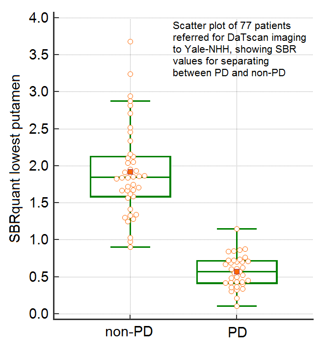
Methods: 401 DaTscan images and their clinical diagnoses were downloaded from PPMI [http://www.ppmi-info.org/]. SBRquant calculated the Striatal Binding Ratio (SBR) separately for the left and right caudate and putamen. The resulting SBRs were evaluated via a receiver operator characteristic’s (ROC) area under the curve (AUC) measure for a range of parameters. The optimum threshold cutoff between HC and PD was found by varying the number of summed transverse slices and positions of the striatal regions of interest (ROI). This optimized SBRquant analyses was applied to 77 patients and ROC analysis yielded the best criteria for separating PD from non-PD patients. The standard of diagnosis of PD versus non-PD was the consensus by the neurologists and nuclear medicine physicians.
Results: The optimized SBRquant analysis of the 77 patients gave SBR values ranging between 0.0 and 4.0, yielding a quantitative measure of the dopaminergic receptor densities in the primarily affected putamen. This measure discriminated the 35 non-PD from the 42 PD patients with a sensitivity and specificity of 97% and 100%.
Conclusions: A fully automated software, SBRquant, has been optimized as a tool to help diagnose PD patients. The optimized parameters applied to a hospital-based cohort yields a discrimination criterion with a nearly perfect correlation with the reported diagnosis between PD and non-PD.
References: I. G. Zubal, M. Early, O. Yuan, D. Jennings, K. Marek, and J. P. Seibyl, “Optimized, automated striatal uptake analysis applied to SPECT brain scans of Parkinson’s disease patients,” J Nucl Med, vol. 48, no. 6, pp. 857-64, Jun, 2007.
To cite this abstract in AMA style:
G. Zubal, S. Tinaz, C. Chow, H. Blumenfeld, E. Louis. Clinical Utility of Optimized Parameters for a Quantitative Analysis of 123I-ioflupane SPECT (DaTscan) Images [abstract]. Mov Disord. 2017; 32 (suppl 2). https://www.mdsabstracts.org/abstract/clinical-utility-of-optimized-parameters-for-a-quantitative-analysis-of-123i-ioflupane-spect-datscan-images/. Accessed April 26, 2025.« Back to 2017 International Congress
MDS Abstracts - https://www.mdsabstracts.org/abstract/clinical-utility-of-optimized-parameters-for-a-quantitative-analysis-of-123i-ioflupane-spect-datscan-images/

