Session Information
Date: Monday, October 8, 2018
Session Title: Parkinson's Disease: Neuroimaging And Neurophysiology
Session Time: 1:15pm-2:45pm
Location: Hall 3FG
Objective: To use radiomics approach based on conventional MRI to classify carriers of LRRK2 mutation among non-manifesting first degree relatives of patients with PD.
Background: The G2019S mutation in LRRK2 gene represents the most common pathogenic genetic mutation identified in Parkinson-disease (PD) worldwide1-2. Healthy carriers of LRRK2 mutations demonstrated changes in brain function, connectivity and reduced DaT-SPECT uptake in comparison to non-carriers. However, structural studies failed to detect any significant differences between healthy carriers and non-carriers. Radiomics analysis is based on the conversion of medical images to high dimensional data set, by extracting a large number of features. This approach was shown to improve prediction over standard radiological assessments especially in oncology applications.
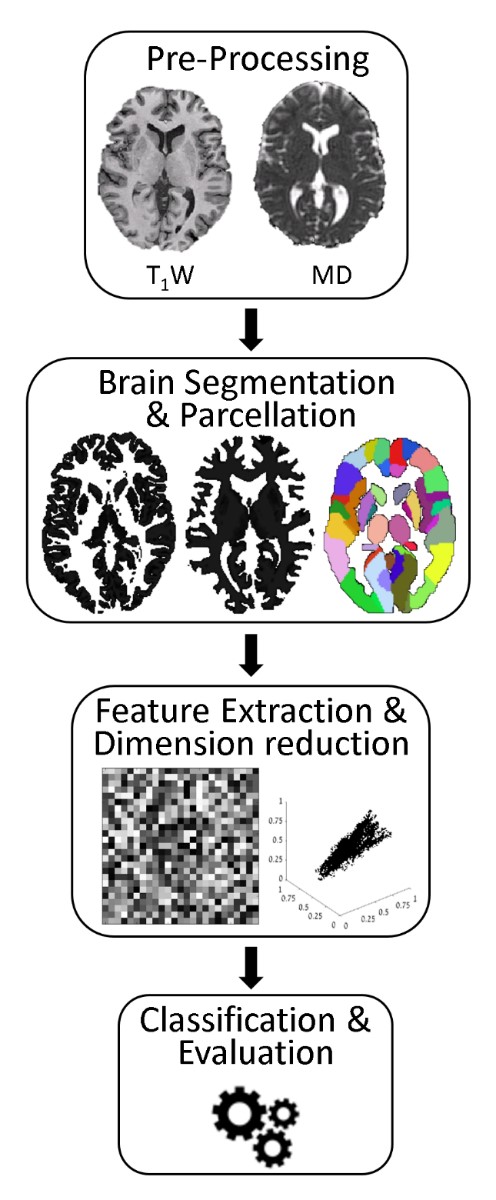
Methods: Subjects and Imaging Data: Seventy-nine healthy Ashkenazi-Jewish subjects were included in this study: 39 non-carriers (4811 y.o) and 40 carriers of the G2019S mutation in LRRK2 gene (4712 y.o). MRI scans were performed on a 3.0T scanner and included high-resolution T1 weighted (T1WI) and diffusion tensor imaging. Image Analysis: Analysis pipeline is illustrated in figure1. Seventeen first and second order statistics parameters were extracted from 100 cortical and subcortical brain regions, based on T1WI and mean diffusivity maps, resulted with a total of 3400 features. Dimensional reduction was performed using principal component analysis and classification was performed using various machine-learning algorithms. Results were evaluated in leave-one subject out manner.
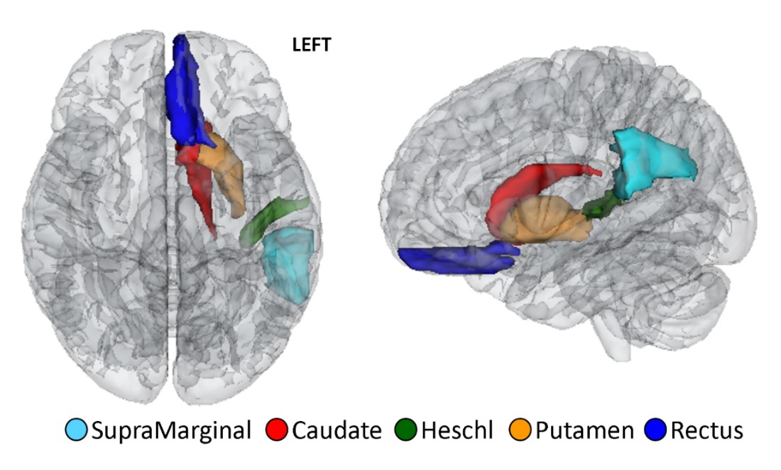
Results: Six features showed significant difference between-group (p<0.01) and were used for classification. Differences were detected only at the left putamen, caudate, straight gyrus, transverse temporal gyrus and the left supra-marginal area (figure2). The best classification results were obtained using a boosted tree classifier, with average accuracy of 80%, sensitivity of 74%, and specificity of 85%.
Conclusions: Radiomics analysis enables to classify healthy carriers of G2019S mutation in the LRRK2 gene from healthy non-carriers, based on differences in several brain regions, known to be related to PD. Studying differences between these two groups may improve our understating regarding the endophenotype of the G2019S mutation in the LRRK2 gene and the pathophysiological mechanism of PD at the prodromal phase of the disease.
References: 1. Farrer, M. et al., Neurology 2005. 2. Orr-Urtreger, A. et al., Neurology 2007.
To cite this abstract in AMA style:
M. Artzi, A. Thaler, A. Orr Urterger, T. Hendler, M. Sela, A. Mirelman, D. Ben Bashat. Classifying carriers of the G2019S mutation in the LRRK2 gene using MRI radiomics analysis [abstract]. Mov Disord. 2018; 33 (suppl 2). https://www.mdsabstracts.org/abstract/classifying-carriers-of-the-g2019s-mutation-in-the-lrrk2-gene-using-mri-radiomics-analysis/. Accessed January 16, 2026.« Back to 2018 International Congress
MDS Abstracts - https://www.mdsabstracts.org/abstract/classifying-carriers-of-the-g2019s-mutation-in-the-lrrk2-gene-using-mri-radiomics-analysis/


