Category: Dystonia: Pathophysiology, Imaging
Objective: The objectives of this work were to create a primate model of dystonia based on striatal infusion of a muscarinic agonist (oxotremorin), and to characterize the clinical phenotype and electrophysiological changes in neuronal activity within the basal ganglia.
Background: Dystonia is a movement disorder characterized by sustained and intermittent muscular co-contractions leading to excessive movements and abnormal postures.
Several genetic models of dystonia have been developed in the past decade in rodents. While they provided interesting insights into the cellular and molecular mechanisms underlying this movement disorder, most of them failed to reproduce an overt phenotype of dystonia except form the model of paroxystic dystonia in the hamster.
A line of evidence supports a specific involvement of the cholinergic system in the pathophysiology of dystonia.
Method: This study was performed on 4 NHPs. Two intracerebral canulas were implanted in the motor part of the striatum of each primate. NaCl- was delivered during control condition and Oxotremorin- M, a non specific cholinergic agonist was used to induce dystonia. Dystonic symptoms were evaluated using a modified version of the BFM scale. Recordings were conducted using high impedance tungsten microelectrodes and targeted the striatum and pallidal regions during control condition and Oxotremorin infusion. Sampling frequency was 20KHz. Firing rate, CV ISI were measured for each sorted unit. Power spectral density of LFP were computed with Welch’s method.
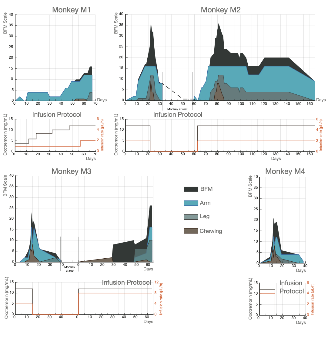
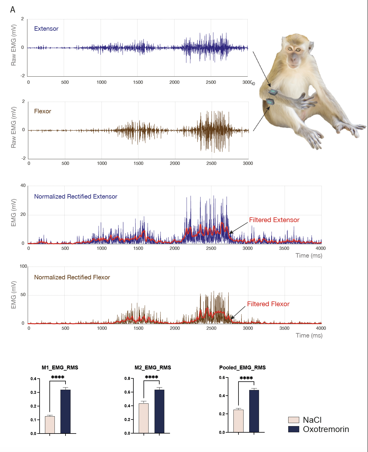
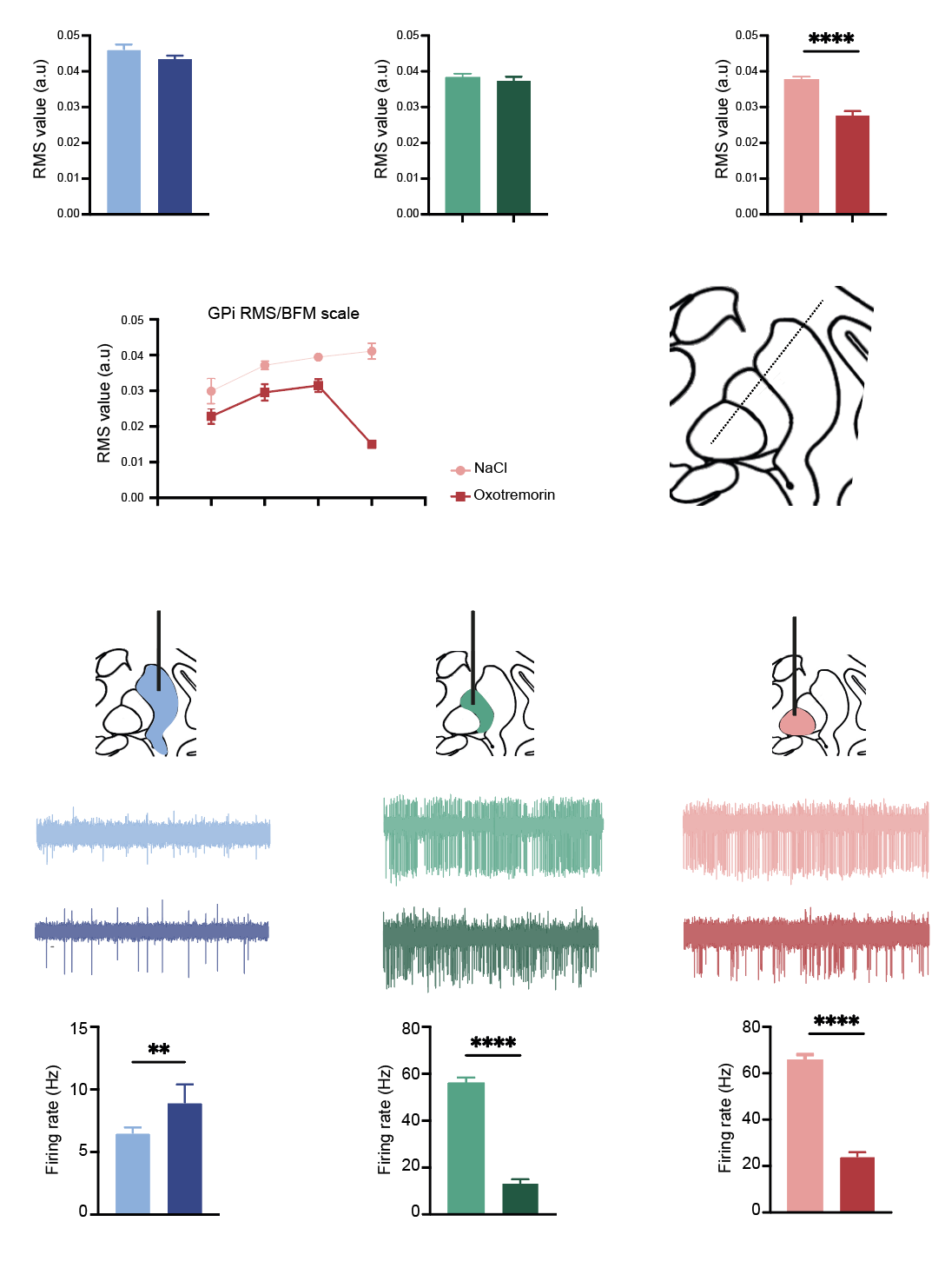
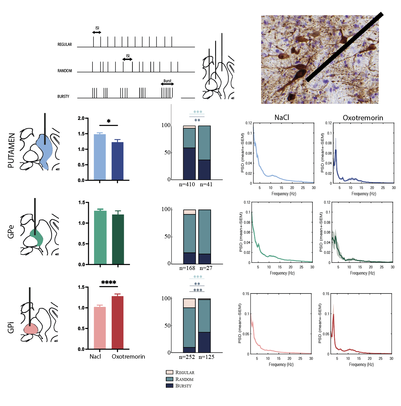
Results: Chronic infusion of non-selective muscarinic agonist (oxotremorin) into the putamen of NHPs led to:(i) abnormal postures and dystonic movements supported by electromyographic recordings; (ii) drastic changes in the firing rate of striatal, external and internal pallidum neurons with firing pattern changes; (iii) changes in oscillatory activity with prominent low frequency activity.
Conclusion: To our knowledge, this is the first symptomatic model of dystonia obtained in non-human primates through chronic infusion of cholinergic agonist into the sensorimotor striatum. Concurrently, we found that neuronal activity changes occurred in the three parts of the lenticular nucleus, namely, the putamen, external and internal globus pallidus suggesting that the dystonic phenotype was associated with a disruption of both the direct and indirect striato-pallidal pathways.
This work will be presented at the AAN 2024 (oral communication)
Dystonic phenotype in NHPs
Elevated EMG RMS values after oxotremorin infusion
Changes in neuronal activity after oxotremorin
Pattern changes and oscillatory activity
To cite this abstract in AMA style:
E. Courtin, B. Ribot, M. Deffains, D. Guehl, P. Burbaud. Chronic striatal cholinergic agonist infusion as a model of dystonia [abstract]. Mov Disord. 2024; 39 (suppl 1). https://www.mdsabstracts.org/abstract/chronic-striatal-cholinergic-agonist-infusion-as-a-model-of-dystonia/. Accessed April 26, 2025.« Back to 2024 International Congress
MDS Abstracts - https://www.mdsabstracts.org/abstract/chronic-striatal-cholinergic-agonist-infusion-as-a-model-of-dystonia/


