Category: Parkinson's Disease: Neuroimaging
Objective: Assess the feasibility of detecting brain iron deposition using quantitative susceptibility mapping (QSM) among patients with Parkinson’s disease (PD) in a community-based clinic setting.
Background: Excessive brain iron accumulation can be found in PD and other neurodegenerative diseases and is thought to enhance free radical formation, contribute to oxidative stress, and result in neuronal death in iron-overloaded cells. Researchers have established brain iron accumulation, as measured by QSM, is associated with cognitive impairment among individuals with PD. Here we report the feasibility of conducting QSM research in a non-academic research center.
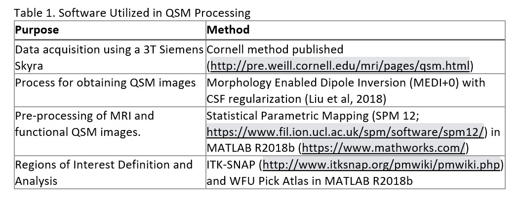
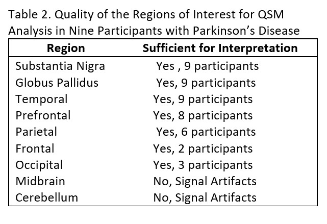
Method: We recruited 9 participants with PD from a community-based clinic to undergo neuroimaging, motor examination (UPDRS-III), depression screening, and a brief cognitive battery. Neuroimages were processed utilizing QSM, guided by previously established methods (Table 1). We examined nine different regions of interest (ROI; Table 2). The study team included a movement disorder neurologist, neuropsychologist, biostatistician, and neuroscience research investigators.
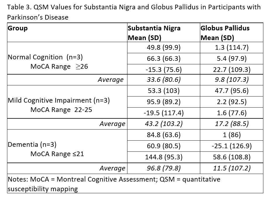
Results: The average age of the participants was 69.3 ± 9.7 (77.8% male; UPDRS-III 30.6 ± 8.8; MoCA = 23.0 ± 5.3; GDS 1.8 ± 2.0). Due to motion artifacts, only 3-4 out of the 9 planned ROI were interpretable, including the substantia nigra, globus pallidus, prefrontal cortex, and temporal regions. Table 3 includes QSM data from the substantia nigra and globus pallidus stratified by the participant’s cognitive status. We discovered a lack of standardization in QSM methods across multiple studies, making cross-study comparisons difficult. Key challenges in processing the images included operating system limitations for existing QSM programs (e.g., our healthcare system does not use Linux) and the lack of a radiologist or neuroradiologist on the team (Table 4).
Conclusion: To our knowledge, there are no studies utilizing QSM to identify brain iron deposition in patients with PD in non-academic research settings; however, we identified several barriers to conducting this research. We recommend further standardization of QSM methods and development of programs/algorithms which can be used across multiple operating systems.
To cite this abstract in AMA style:
B. Kashyap, K. Wyman-Chick, L. Hanson, S. Gustafson, S. Sherman, W. Frey, Ii, J. Johnson. Challenges in the measurement of brain iron deposition in Parkinson’s disease in non-academic research settings. [abstract]. Mov Disord. 2023; 38 (suppl 1). https://www.mdsabstracts.org/abstract/challenges-in-the-measurement-of-brain-iron-deposition-in-parkinsons-disease-in-non-academic-research-settings/. Accessed April 20, 2025.« Back to 2023 International Congress
MDS Abstracts - https://www.mdsabstracts.org/abstract/challenges-in-the-measurement-of-brain-iron-deposition-in-parkinsons-disease-in-non-academic-research-settings/


