Session Information
Date: Monday, September 23, 2019
Session Title: Rare Genetic and Metabolic Diseases
Session Time: 1:45pm-3:15pm
Location: Les Muses Terrace, Level 3
Objective: Cerebrontendinous xanthomatosis(CTX) is a treatable, autosomal recessive, lipid storage disorder disease. Herein, we present a case of CTX. It aims to refresh and improve the Parkinson’s specialists’ understanding of the disease.
Background: A 40-year-old man was referred to our wards with a 10-year history of progressive gait difficulty and imbalance, bradykinesia and involuntary shaking of both hands. There was a history of bilateral cataracts and epileptic seizures. He was ever given “Madopar” for one year. But there was no effect.
Method: We collected neurologic examination, blood indicators, cerebrospinal fluid examination, steroid spectrum, abdominal ultrasound, EEG, EMG, CT, MRI, SWI, Achilles tendon pathology, gene sequencing, FDG-PET, AV133-PET. Follow-up was performed after treatment.
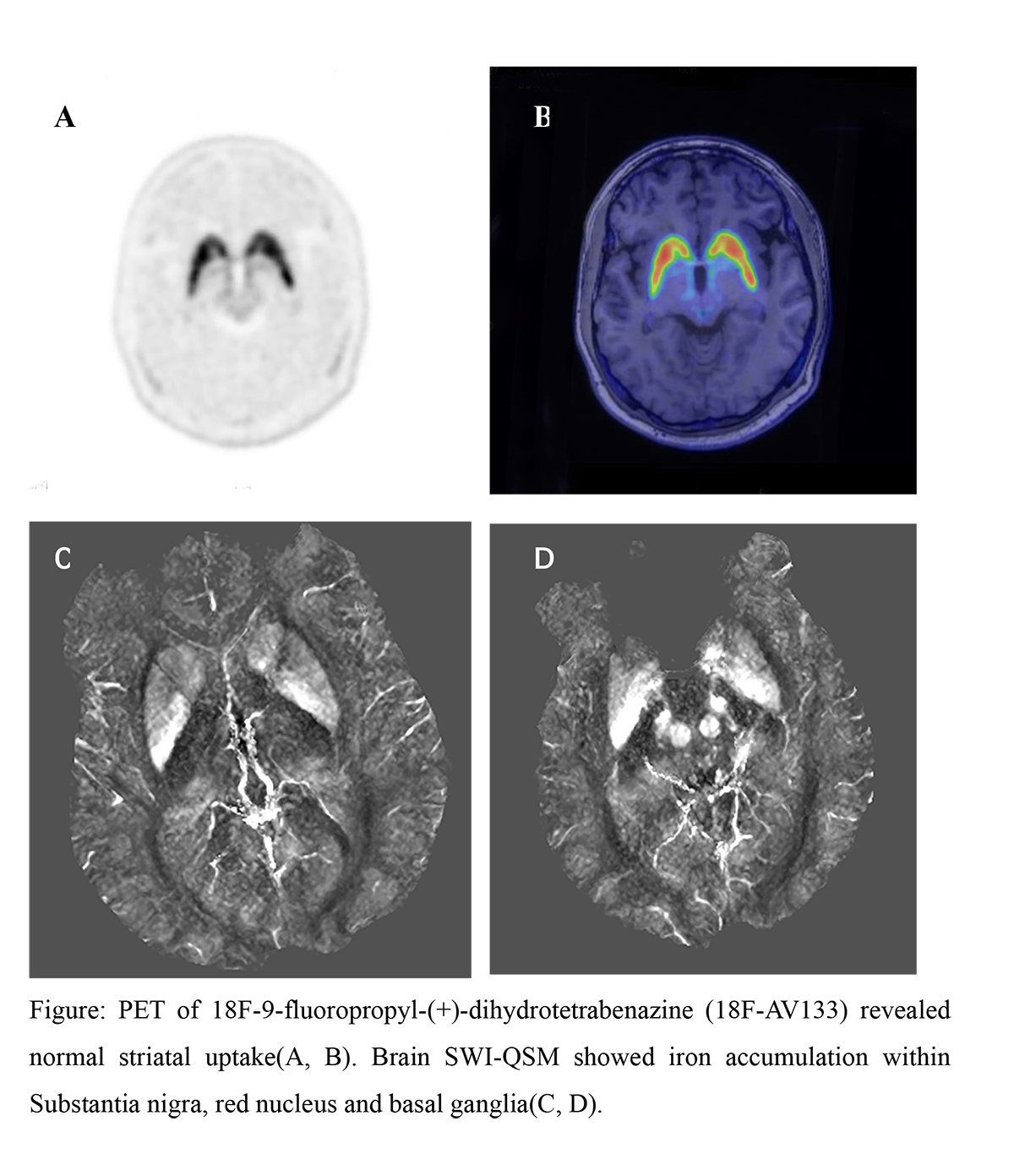
Results: Neurologic examination indicates pyramidal and extrapyramidal damage. Brain MRI shows white matter degeneration, and the symmetry abnormal signal of the cerebellar dentate nucleus. Positron Emission Tomography (PET) of vesicular monoamine transporter 2 (VMAT2) revealed normal striatal uptake (Figure A, B). Brain SWI quantitative susceptibility mapping shows iron accumulation within bilateral substantia nigra, red nucleus and basal ganglia (Figure C, D). Achilles tendon pathology suggests multifocal xanthoma cells and multiple cholesterol crystals to make. Gene sequencing indicated two heterozygous mutations in cyp27a1.The patient improved after oral treatment of ursodeoxycholic acid for 2 weeks, but no improvement when he changed to chenodeoxycholic acid (CDCA) later at 12 weeks.
Conclusion: Parkinsonism is rare and can be as the only neurological manifestation in CTX[1]. Early diagnosis and start at an early age can reverse or even prevent the development of neurologic symptoms in CTX[2]. Parkinsonism seemed as a treatment-resistant feature of CTX[2,3].Functional dopaminergic imaging showed presynaptic denervation in some patients[1,2]. Parkinsonism can be regarded as advanced disease manifestations and are often under underestimated. The mechanism of Parkinsonism in CTX is not clear. In this paper, brain SWI image suggests iron deposition in substantia nigra, red nucleus and basal ganglia, which may be related to the mechanism of Parkinsonism and unreported before.
References: 1.Mignarri A, Falcini M, Vella A, etal. Parkinsonism as neurological presentation of late-onset cerebrotendinous xanthomatosis. Parkinsonism Relat Disord. 2012 Jan;18(1):99-101. doi: 10.1016/j.parkreldis.2011.06.004. Epub 2011 Jul 20. 2.Bianca M.L. Stelten, MD, Hidde H. Huidekoper, MD, PhD, etal. Long-term treatment effect in cerebrotendinous xanthomatosis depends on age at treatment start. Neurology 2019;92:1-13. doi:10.1212/WNL.0000000000006731. 3. Movement disorders in cerebrotendinous xanthomatosis. Parkinsonism Relat Disord. 2018 Jul 19. pii: S1353-8020(18)30310-9. doi:10.1016/j.parkreldis.2018.07.006. [Epub ahead of print]
To cite this abstract in AMA style:
J. Li, S. Liu, E. Xu, H. Qiao, W. Mao, P. Chan. Cerebrotendinous Xanthomatosis presenting Parkinsonism with bilateral iron accumulation in the basal ganglia [abstract]. Mov Disord. 2019; 34 (suppl 2). https://www.mdsabstracts.org/abstract/cerebrotendinous-xanthomatosis-presenting-parkinsonism-with-bilateral-iron-accumulation-in-the-basal-ganglia/. Accessed April 22, 2025.« Back to 2019 International Congress
MDS Abstracts - https://www.mdsabstracts.org/abstract/cerebrotendinous-xanthomatosis-presenting-parkinsonism-with-bilateral-iron-accumulation-in-the-basal-ganglia/

