Category: Rare Genetic and Metabolic Diseases
Objective: We present a case series of cerebellar ataxia due to hypomyelinating leukodystrophies (HLDs).
Background: HLDs are a group of disorders arising from primary deficit in myelin development, being characterized radiologically by near normal T1 and T2 hyperintensity of the cerebral white matter [1]. While most of them present in infancy, late onset phenotypes have also been reported. Movement disorders in the form of cerebellar ataxia, dystonia, tremor and choreoathetosis can be part of the varied clinical manifestations of HLDs, along with associated morphological features like dysmorphism, hypogonadism and dental abnormalities [2,3].
Method: We screened the cerebellar ataxia patients attending the movement disorder clinic at Institute of Neurosciences Kolkata (I-NK) from 1st Jan 2021 to 31st Dec 2022. They were stratified as per possible etiology. Among them, we selected 6 patients with HLD based on their clinical and radiological findings.
Results: Out of these 6 patients with HLD, 3 had Pelizaeus–Merzbacher like disease 1 (PMLD1) from pathogenic variants in the GJC2 gene and 2 had POLR3 spectrum disorder, 1 with POLR3A and the other with POLR3K variants. 1 patient had DARS1 pathogenic variant giving rise to Hypomyelination with brainstem and spinal cord involvement with leg spasticity (HBSL). Cerebellar ataxia along with pyramidal signs were prominent in all of them and the age of presentation varied from 3 to 26 years (mean 13.66 ± 9.21) with mean disease duration of 5 ± 3.5 years. Dystonia was present in 5 of them (focal in HBSL, segmental in POLR3A and generalized in PMLD1). Nystagmus (elliptical pendular and upbeat) was striking in patients of PMLD1. Dental abnormalities (malocclusion of teeth, persistent natal teeth) [figure1] and hypogonadism with small testis were present in POLR3 variants.
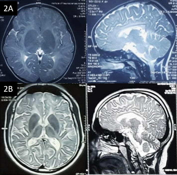
MRI brain revealed diffuse white matter T2 hyperintensities with near normal T1 intensity in all 6 patients. Relative T2 hypointensities of the globus pallidus and ventrolateral thalamus noted in POLR3 variants. Cerebellar atrophy was present in all patients while thin corpus callosum was evident in PMLD1 and in POLR3 variants [figure2]. The patient with HBSL showed T2 hyperintensity in superior cerebellar peduncle and dorsal column of spinal cord.
Conclusion: HLDs should be kept in differentials of spastic cerebellar ataxia. Associated morphological and imaging features provide vital clues to diagnose them.
References: [1] Wolf NI, Ffrench-Constant C, van der Knaap MS. Hypomyelinating leukodystrophies – unravelling myelin biology. Nat Rev Neurol. 2021 Feb;17(2):88-103. doi: 10.1038/s41582-020-00432-1.
[2] Pouwels PJ, Vanderver A, Bernard G, Wolf NI, Dreha-Kulczewksi SF, Deoni SC, Bertini E, Kohlschütter A, Richardson W, Ffrench-Constant C, Köhler W, Rowitch D, Barkovich AJ. Hypomyelinating leukodystrophies: translational research progress and prospects. Ann Neurol. 2014 Jul;76(1):5-19. doi: 10.1002/ana.24194.
[3] Di Bella D, Magri S, Benzoni C, Farina L, Maccagnano C, Sarto E, Moscatelli M, Baratta S, Ciano C, Piacentini SHMJ, Draghi L, Mauro E, Pareyson D, Gellera C, Taroni F, Salsano E. Hypomyelinating leukodystrophies in adults: Clinical and genetic features. Eur J Neurol. 2021 Mar;28(3):934-944. doi: 10.1111/ene.14646.
To cite this abstract in AMA style:
J. Ganguly, J. Sinha, P. Basu, M. Tiwari, H. Kumar. Cerebellar ataxia in Hypomyelinating Leukodystrophies: A case series [abstract]. Mov Disord. 2023; 38 (suppl 1). https://www.mdsabstracts.org/abstract/cerebellar-ataxia-in-hypomyelinating-leukodystrophies-a-case-series/. Accessed April 26, 2025.« Back to 2023 International Congress
MDS Abstracts - https://www.mdsabstracts.org/abstract/cerebellar-ataxia-in-hypomyelinating-leukodystrophies-a-case-series/


