Session Information
Date: Thursday, June 8, 2017
Session Title: Dystonia
Session Time: 1:15pm-2:45pm
Location: Exhibit Hall C
Objective: To explore the relationship between brain morphology, clinical effects of bilateral pallidal stimulation (GPi DBS) and intracortical inhibition of the motor cortex in patients with dystonia.
Background: Dystonia patients have trouble suppressing unwanted movement that is potentially related to less effective intracortical inhibition, which can be restored with GPi DBS. However, the treatment efficacy varies and the benefit increases relatively slowly, suggesting induction of slow plastic processes in regions involved with motor control.
Methods: We examined 19 patients (mean age 48±(SD)18 years) with cervical (N=7) or generalized dystonia (N=12) of various origin by chronic GPi DBS for 57±28 months. Voxel-based morphometry of postoperative T1-weighted images (MPRAGE, 1x1x1 mm) was calculated for gray matter (GM) density in each voxel using CAT12/SPM12 software in every patient and compared with 20 matched controls. Paired TMS with subthreshold conditioning stimulus followed by a supratreshold testing stimulus were applied to the motor cortex to elicit short-latency intracortical inhibition (SICI) of the motor evoked potential. The clinical effect of GPi DBS was expressed as a change in the dystonic score (BFMDS or TWSTRS) between actual GPi DBS ON condition and the preoperative state.
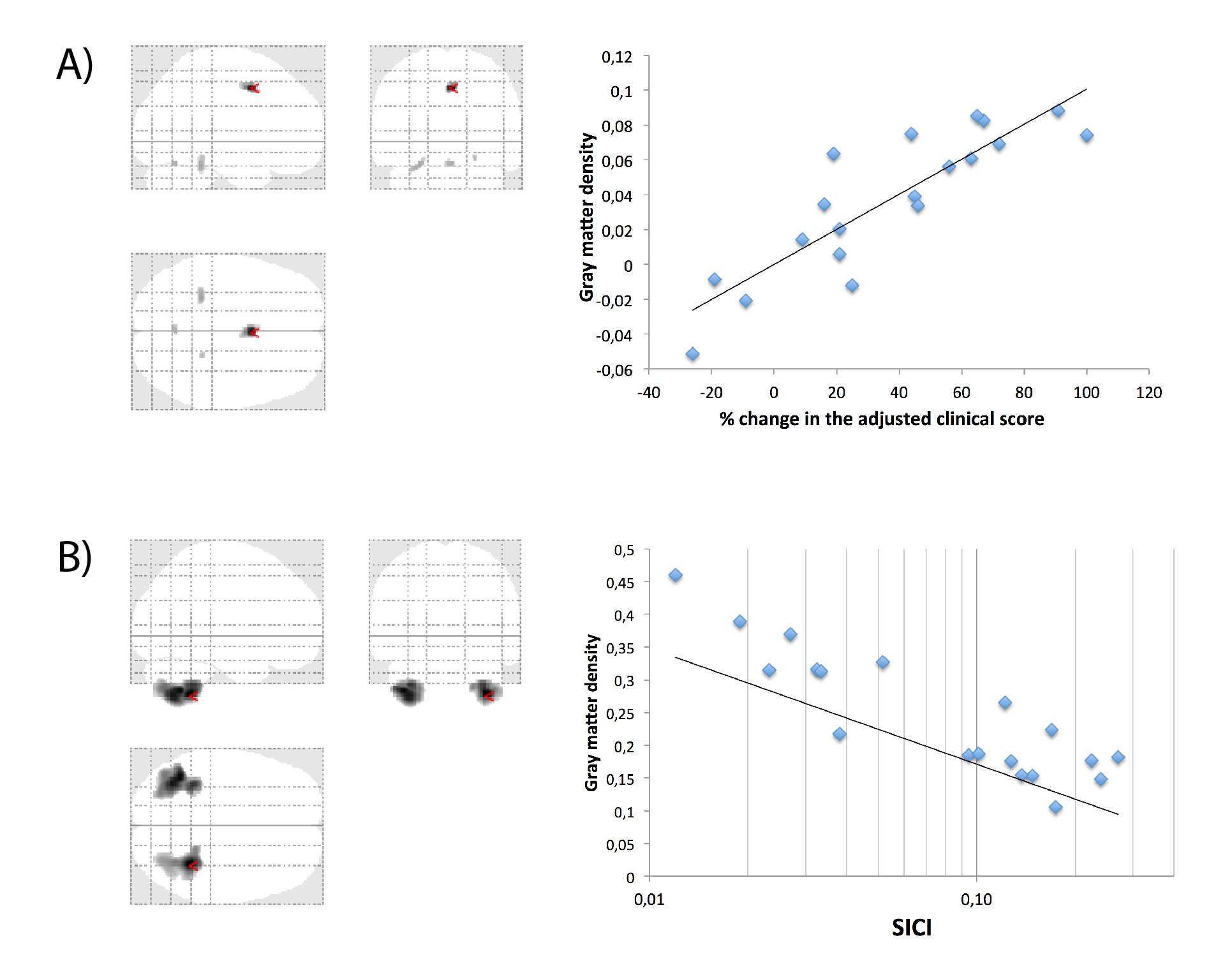
Results: Dystonia patients showed increased GM density in the supplementary motor area (SMA) and middle cingulate in comparison with healthy controls (p<0.05 corrected). The GM density in this region positively correlated with the clinical effect of GPi DBS (p<0.001)(Fig. 1A). The SICI was lower in patients than in controls regardless of the ON and OFF conditions (p<0.001) and its mean amplitude correlated with GM density in both cerebellar hemispheres (p<0.05 corrected)(Fig. 1B).[figure1]
Conclusions: Brain changes of chronically GPi DBS treated patients possibly reflect “hardwire” rebuilding of motor regions associated with functional improvement. Cortically, they showed that the SMA hypertrophy was quantitatively related to the clinical benefit suggesting a compensatory mechanism. Subcortically, they showed hypertrophy of the cerebellar hemispheres growing with the effectiveness of intracortical inhibition of the primary motor cortex which possibly allows better motor control. Supported by the grants GACR 16-13323S and PRVOUK P26/LF1/4.
To cite this abstract in AMA style:
R. Jech, A. Fečíková, V. Čejka, V. Čapek, D. Šťastná, F. Růžička, D. Urgošík, E. Růžička. Brain volume changes in relation to intracortical inhibition and clinical benefit of pallidal stimulation in dystonia: a combined VBM and TMS study. [abstract]. Mov Disord. 2017; 32 (suppl 2). https://www.mdsabstracts.org/abstract/brain-volume-changes-in-relation-to-intracortical-inhibition-and-clinical-benefit-of-pallidal-stimulation-in-dystonia-a-combined-vbm-and-tms-study/. Accessed December 27, 2025.« Back to 2017 International Congress
MDS Abstracts - https://www.mdsabstracts.org/abstract/brain-volume-changes-in-relation-to-intracortical-inhibition-and-clinical-benefit-of-pallidal-stimulation-in-dystonia-a-combined-vbm-and-tms-study/

