Session Information
Date: Wednesday, September 25, 2019
Session Title: Neuroimaging
Session Time: 1:15pm-2:45pm
Location: Les Muses Terrace, Level 3
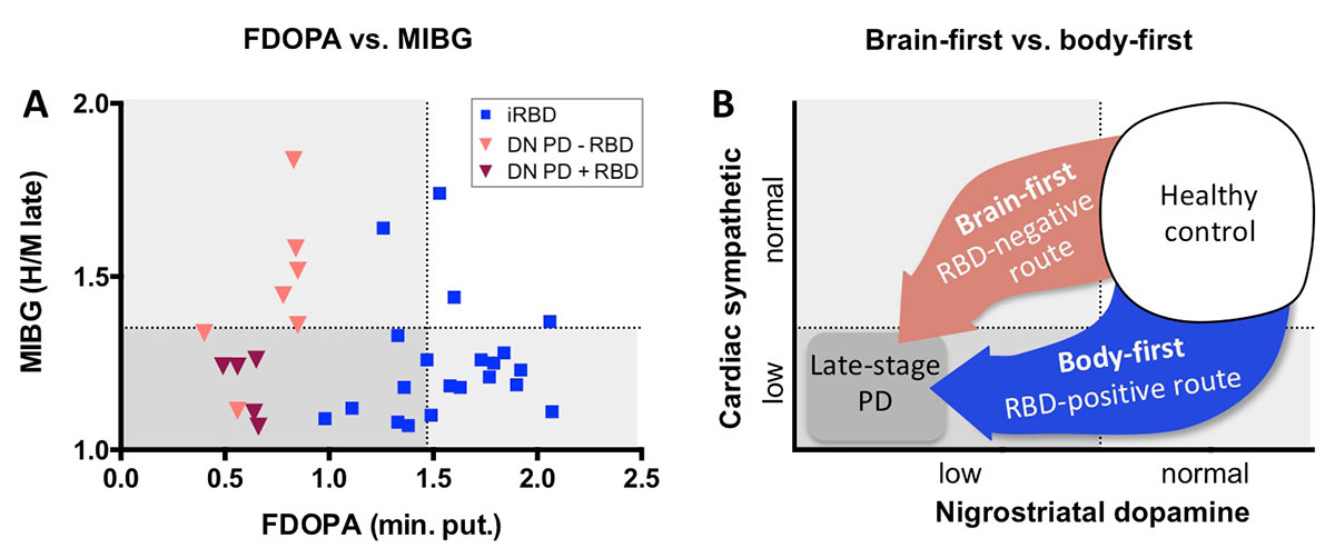
Objective: In a multi-modality imaging study, we test the hypothesis that Parkinson’s disease (PD) can be divided into two strikingly distinct phenotypes: (1) PD patients, in whom RBD appears before diagnosis, is a “body-first” phenotype characterized by early, severe damage to the autonomic nervous system, while the dopamine system is still mostly intact. (2) RBD-negative PD is a “brain-first” phenotype, characterized by marked, early damage to the nigrostriatal dopamine system, while the peripheral autonomic system is relatively intact.
Background: In a multi-modality imaging study, we recently demonstrated that RBD patients show full-blown damage to the parasympathetic and sympathetic nervous systems, and to the locus coeruleus, which were at the level of diagnosed PD. Yet, 71% of RBD patients had normal dopaminergic storage capacity measured with FDOPA PET (1). In contrast, up to 50% of H&Y stage I PD patients with unknown RBD-status still have normal cardiac sympathetic innervation measured by MIBG scintigraphy (2, 3). By extrapolation, these H&Y-I PD patients must be mostly RBD-negative.
Method: We aim to include 100 de novo, untreated PD patients into this cohort. All patients have polysomnography and 11C-donepezil PET/CT, 123I-MIBG scintigraphy, neuromelanin MRI, and 18F-FDOPA PET – measures of parasympathetic, sympathetic, locus coeruleus, and the nigro-striatal innervation, respectively. Olfaction, cognitive, motor, and non-motor status is assessed. The data is compared to in-house data from healthy controls, idiopathic RBD (iRBD) and manifest PD.
Results: Here, we present FDOPA and MIBG data from the first 12 de novoPD patients (DN-RBD); 7 RBD-negative, 5 RBD-positive and compare to iRBD data [figure1]. All DN-RBD had pathological FDOPA PET scans, but only 1/7 had manifestly pathological MIBG scan. In contrast, 17/21 iRBD cases had pathological MIBG scan, but only 7/21 showed pathological FDOPA scans (p=0.003).
Conclusion: These initial data supports the hypothesis. Most de novo, RBD-negative PD patients have developed marked dopaminergic loss in the absence of marked cardiac sympathetic loss. We propose that the initial Lewy pathology in RBD-negative PD originates in the brainstem with secondary brain-to-periphery spreading, whereas the initial pathology in iRBD originates in the periphery (gut) and then spreads to the brainstem.
References: 1. Knudsen K, Fedorova TD, Hansen AK, Sommerauer M, Otto M, Svendsen KB, Nahimi A, Stokholm MG, Pavese N, Beier CP, Brooks DJ, Borghammer P. Lancet Neurology 2018: 17(7):618-28. 2. Kashihara K, Imamura T, Shinya T. Cardiac 123I-MIBG uptake is reduced more markedly in patients with REM sleep behavior disorder than in those with early stage Parkinson’s disease. Park Relat Disord 2010: 16:252-53. 3. Nagayama H, Hamamoto M, Nagashima J, Katayama Y. Reliability of MIBG myocardial scintigraphy in the diagnosis of Parkinson’s disease. J Neurol Neurosurg Psychiatry 2005; 76:249-51.
To cite this abstract in AMA style:
J. Horsager, K. Knudsen, K. Andersen, C. Skjærbæk, T. Fedorova, J. Geday, J. Kraft, E. Bech, E. Danielsen, M. Møller, N. Pavese, D. Brooks, P. Borghammer. “Brain-first” vs. “body-first” Parkinson’s disease is determined by RBD-status – a multi-modality imaging study [abstract]. Mov Disord. 2019; 34 (suppl 2). https://www.mdsabstracts.org/abstract/brain-first-vs-body-first-parkinsons-disease-is-determined-by-rbd-status-a-multi-modality-imaging-study/. Accessed December 29, 2025.« Back to 2019 International Congress
MDS Abstracts - https://www.mdsabstracts.org/abstract/brain-first-vs-body-first-parkinsons-disease-is-determined-by-rbd-status-a-multi-modality-imaging-study/

