Category: Parkinson's Disease: Neuroimaging
Objective: To identify subthalamic clusters that predict maximum improvement in bradykinesia, rigidity and tremor. To map-out the cortical fingerprint, which predict maximum efficacy.
Background: Subthalamotomy using transcranial Magnetic Resonance-guided Focused Ultrasound (MRgFUS) has become a therapeutic approach that can efficiently improve all motor features in patients with Parkinson’s disease (PD). However, a more detailed characterization of the targeted network and its cortical terminals that drive the clinical outcome is needed, in order to understand its therapeutic mechanism.
Method: A total of 39 PD patients undergoing MRgFUS-subthalamotomy were scanned with high resolution MRI 24 hours after treatment. Motor scores of the Unified Parkinson Disease Rating Scale [UPDRS-III] were computed at baseline and at 4-month follow-up after treatment. Manual segmentation and voxel-based statistical analysis of lesions was used to identify significant treatment clusters. Publicly available tractography-based connectome data was employed to identify cortical connectivity patterns associated with treatment efficacy.
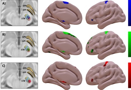
Results: Results. Greater improvement in bradykinesia was explained by more anterior lesions along the anteroposterior axis. Anti-tremor effect increases as the lesion impacts more within the posterior-lateral region of the motor STN. The rigidity effective cluster lay in between and overlapped with both STN subregions. Cortical connectivity to supplementary motor area (SMA) was predictive of improvement in bradykinesia (figure 1A); whilst that to primary motor area (M1) was predictive of higher improvement in tremor (figure 1C). Connectivity to SMA and prefrontal cortices was predictive of improvement in rigidity (figure 1B).
[figure1]
Conclusion: These data suggest that maximum improvement in bradykinesia, tremor and rigidity induced by subthalamotomy largely rely on the location of lesions and the concomitant effect on cortical connectivity patterns, particularly to M1 and SMA.
To cite this abstract in AMA style:
R. Rodriguez-Rojas, JU. Mañez-Miro, JA. Pineda-Pardo, R. Martinez-Fernandez, M. Del Alamo, JA. Obeso. Beyond the subthalamic nucleus: tractography patterns of Magnetic Resonance-guided Focused Ultrasound subthalamotomy [abstract]. Mov Disord. 2022; 37 (suppl 2). https://www.mdsabstracts.org/abstract/beyond-the-subthalamic-nucleus-tractography-patterns-of-magnetic-resonance-guided-focused-ultrasound-subthalamotomy/. Accessed December 30, 2025.« Back to 2022 International Congress
MDS Abstracts - https://www.mdsabstracts.org/abstract/beyond-the-subthalamic-nucleus-tractography-patterns-of-magnetic-resonance-guided-focused-ultrasound-subthalamotomy/

