Category: Parkinson's Disease: Neuroimaging
Objective: To investigate the associations between the choroid plexus (ChP) and protein indicators of Parkinson’s Disease (PD).
Background: The ChP is the primary organ that produces cerebrospinal fluid (CSF), which is the carrier of the glymphatic system. Therefore, the structure and the function of ChP may be closely associated with the clearance of protein indicators in PD. Previous studies, however, either disregarded the choroid plexus as an interfering organ or were constrained by inaccuracies in the ChP segmentation. [1][2]. Therefore, an in-house ChP segmentation pipeline was developed based on the deep learning model of 3D UNet to investigate the alternations in the ChP among PD patients.
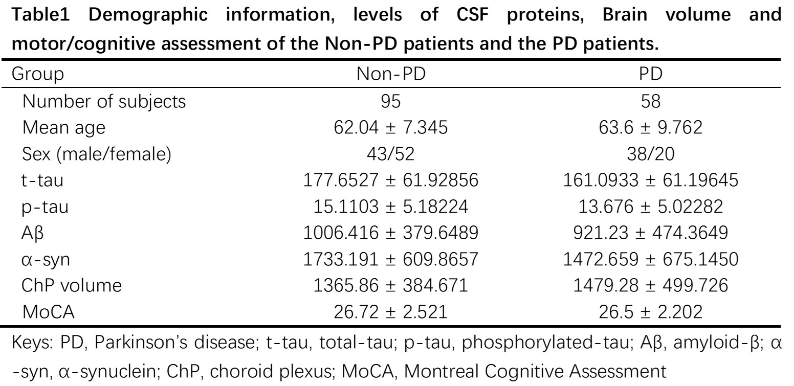
Method: 95 non-PD patients (including healthy controls and prodromal) and 58 PD patients were selected from the PPMI dataset based on the availability of T1-weighted MR images and protein indicators, Table 1. ChP was segmented based on the in-house pipeline and its volume was calculated. The relationships between ChP volume and CSF proteins, including t-tau, p-tau, Aβ and α-syn, were examined by controlling for age and gender using multivariate analysis in the SPSS. Additionally, the relationships between ChP volume and CSF proteins were compared among cognitive impairment and non-cognitive impairment PD patients according to Montreal Cognitive Assessment (MoCA) scores, using a threshold of 25.
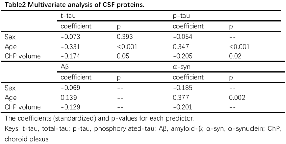
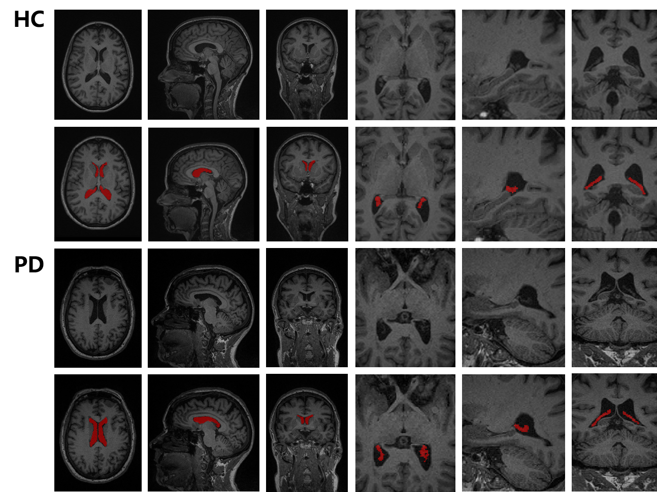
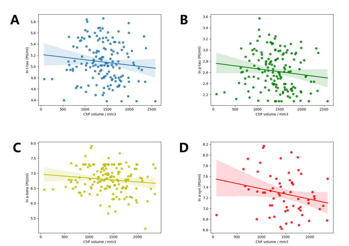
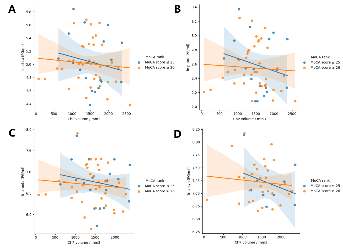
Results: The in-house pipeline provided accurate ChP segmentation, Figure 1. Compared to the Non-PD group, PD patients showed 8.30% larger ChP volume (p=0.117). A significant and negative correlation between ChP volume and p-tau was found (p=0.02) while the marginal correlation was found between ChP volume and t-tau (p=0.05), Figure 2 and Table 2. PD patients with cognitive impairment (MoCA scores below 25) showed lower negative correlations between ChP volume and CSF proteins compared to the non-cognitive impairment group (t-tau: -0.175 vs -0.088, p-tau: -0.218 vs -0.061, Aβ: -0.182 vs -0.094, and α-syn: -0.32 vs -0.1), Figure 3, which may suggest the dysfunction of the glymphatic system..
Conclusion: The present study revealed a negative relationship between choroid plexus volume and both p-tau and t-tau, and the correlation coefficients decrease as PD progresses. -· 123
Table1 Basic information of patients.
Table2 Multivariate analysis of CSF proteins.
Figure 1. ChP segmentation
Figure 2. ChP volume and protein indicators.
Figure 3. ChP volume and protein in PD patients.
Figure illustration
References: [1] L. Zhao, X. Feng, C. H. Meyer and D. C. Alsop, “Choroid Plexus Segmentation Using Optimized 3D U-Net,” 2020 IEEE 17th International Symposium on Biomedical Imaging (ISBI), Iowa City, IA, USA, 2020, pp. 381-384, doi: 10.1109/ISBI45749.2020.9098443.
[2] Tadayon E, Pascual-Leone A, Press D, Santarnecchi E; Alzheimer’s Disease Neuroimaging Initiative. Choroid plexus volume is associated with levels of CSF proteins: relevance for Alzheimer’s and Parkinson’s disease. Neurobiol Aging. 2020 May;89:108-117. doi: 10.1016/j.neurobiolaging.2020.01.005. Epub 2020 Jan 16. PMID: 32107064; PMCID: PMC9094632.
To cite this abstract in AMA style:
Z. Wang, J. Li, C. Cai, C. Jiang, L. Zhao. Associations between Choroid Plexus and tau in Parkinson’s Disease [abstract]. Mov Disord. 2024; 39 (suppl 1). https://www.mdsabstracts.org/abstract/associations-between-choroid-plexus-and-tau-in-parkinsons-disease/. Accessed April 20, 2025.« Back to 2024 International Congress
MDS Abstracts - https://www.mdsabstracts.org/abstract/associations-between-choroid-plexus-and-tau-in-parkinsons-disease/

