Category: Surgical Therapy: Parkinson's Disease
Objective: To characterize globus pallidus internus (GPi) deep brain stimulation (DBS) active contact location at one-year post-op for Parkinson’s disease (PD) patients at our center.
Background: Traditionally, the posteroventral GPi has been used as a DBS target for PD. Recently, other locations within the GPi have been shown to be efficacious. To glean a better understanding of other potentially efficacious GPi subregions, we reviewed our institution’s active contact location and outcome at one-year in PD patients who underwent GPi DBS.
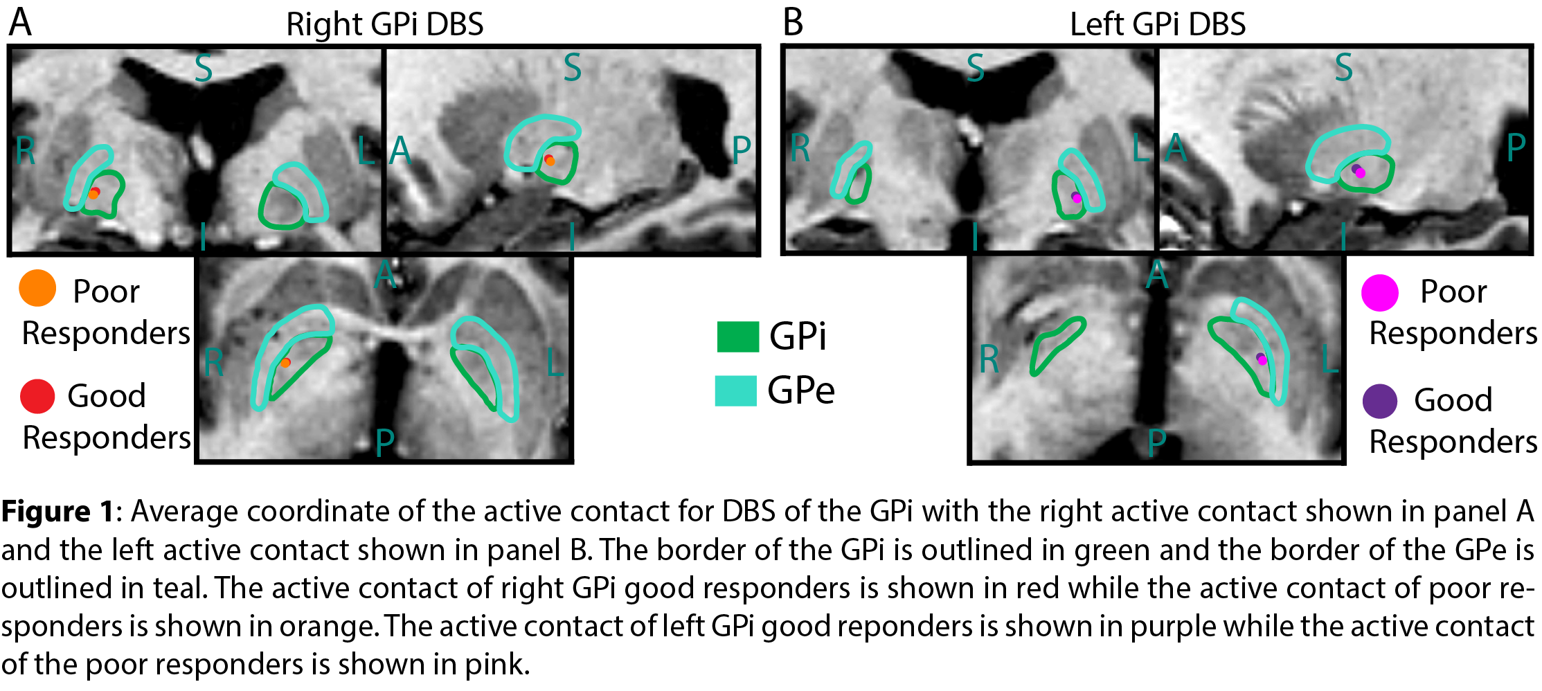
Method: Records of PD patients who underwent GPi DBS at VUMC from 2011-2021 were reviewed. 82 patients with PD with at least one-year neurology follow-up were identified. Clinic notes by the treating neurologist one-year post-DBS placement were reviewed to determine the optimized DBS contact. Clinical improvement was rated with a score of 1-4 (no, minimal, moderate, marked improvement, respectively). Good responders were defined as patients with a score of 3 or 4 and poor responders were defined as patients with a score of 1 or 2. A functional brain atlas with normalized imaging space (Cranial Vault Explorer) was used to visualize active contact locations. A 2-sample t-test was used to evaluate significant differences of active contact locations between the two groups.
Results: Among the patients reviewed, average active contact coordinates relative to the midcommissural point in atlas space were as follows: good responders left GPi (22.15mm lateral, 3.70mm anterior, 1.10mm inferior), good responders right GPi (22.14mm lateral, 5.06mm anterior, 1.04mm superior), poor responders left GPi (22.56mm lateral, 3.11mm anterior, 1.39mm inferior), poor responders right GPi (22.43mm lateral, 4.74mm anterior, 0.73mm superior). There was no statistically significant difference in average contact location between good and poor responders (p-values: Left Lateral 0.437, AP 0.326, vertical 0.740; Right lateral 0.543, AP 0.526, vertical 0.746).
Conclusion: The active contact location in PD patients undergoing GPi DBS was located dorsally near the GPi border relative to the typical posteroventral lead tip location at our center. There was no statistically significant difference in active contact location between good and poor responders, emphasizing the importance of patient selection and individualized programming.
References: Holland MT, Jiao J, Mantovani A, Anderson S, Mitchell KA, Safarpour D, Burchiel KJ. Identifying the therapeutic zone in globus pallidus deep brain stimulation for Parkinson’s disease. J Neurosurg. 2022 Jul 22;138(2):329-336. doi: 10.3171/2022.5.JNS22152. PMID: 35901683.
D’Haese PF, Pallavaram S, Li R, Remple MS, Kao C, Neimat JS, Konrad PE, Dawant BM. CranialVault and its CRAVE tools: a clinical computer assistance system for deep brain stimulation (DBS) therapy. Med Image Anal. 2012 Apr;16(3):744-53. doi: 10.1016/j.media.2010.07.009. Epub 2010 Aug 1. PMID: 20732828; PMCID: PMC3021628.
To cite this abstract in AMA style:
D. Schumacher, D. Paulo, D. Doss, S. Narasimhan, L. Rui, D. Benoit, D. Englot, A. Giritharan. Assessing the optimal pallidal target in deep brain stimulation for Parkinson’s disease: A single-center retrospective review [abstract]. Mov Disord. 2023; 38 (suppl 1). https://www.mdsabstracts.org/abstract/assessing-the-optimal-pallidal-target-in-deep-brain-stimulation-for-parkinsons-disease-a-single-center-retrospective-review/. Accessed February 6, 2026.« Back to 2023 International Congress
MDS Abstracts - https://www.mdsabstracts.org/abstract/assessing-the-optimal-pallidal-target-in-deep-brain-stimulation-for-parkinsons-disease-a-single-center-retrospective-review/

