Category: Parkinson's Disease: Neuroimaging
Objective: We aimed to use diffusion MRI quantitative maps as an objective, non-invasive measure to assess for gait disturbances in Parkinson’s Disease (PD).
Background: PD presents with varying gait symptoms. These range from no gait disturbance (NGD) to gait disturbance (GD) to freezing-of-gait (FOG). GD includes slowing gait, shuffling, and instability, while FOG is the inability to take a step, potentially leading to falls. Quantitative maps like diffusion tensor imaging (DTI), diffusion kurtosis imaging (DKI), and neurite orientation dispersion and density imaging (NODDI) have the potential to be used as an objective assessment for gait disturbances.
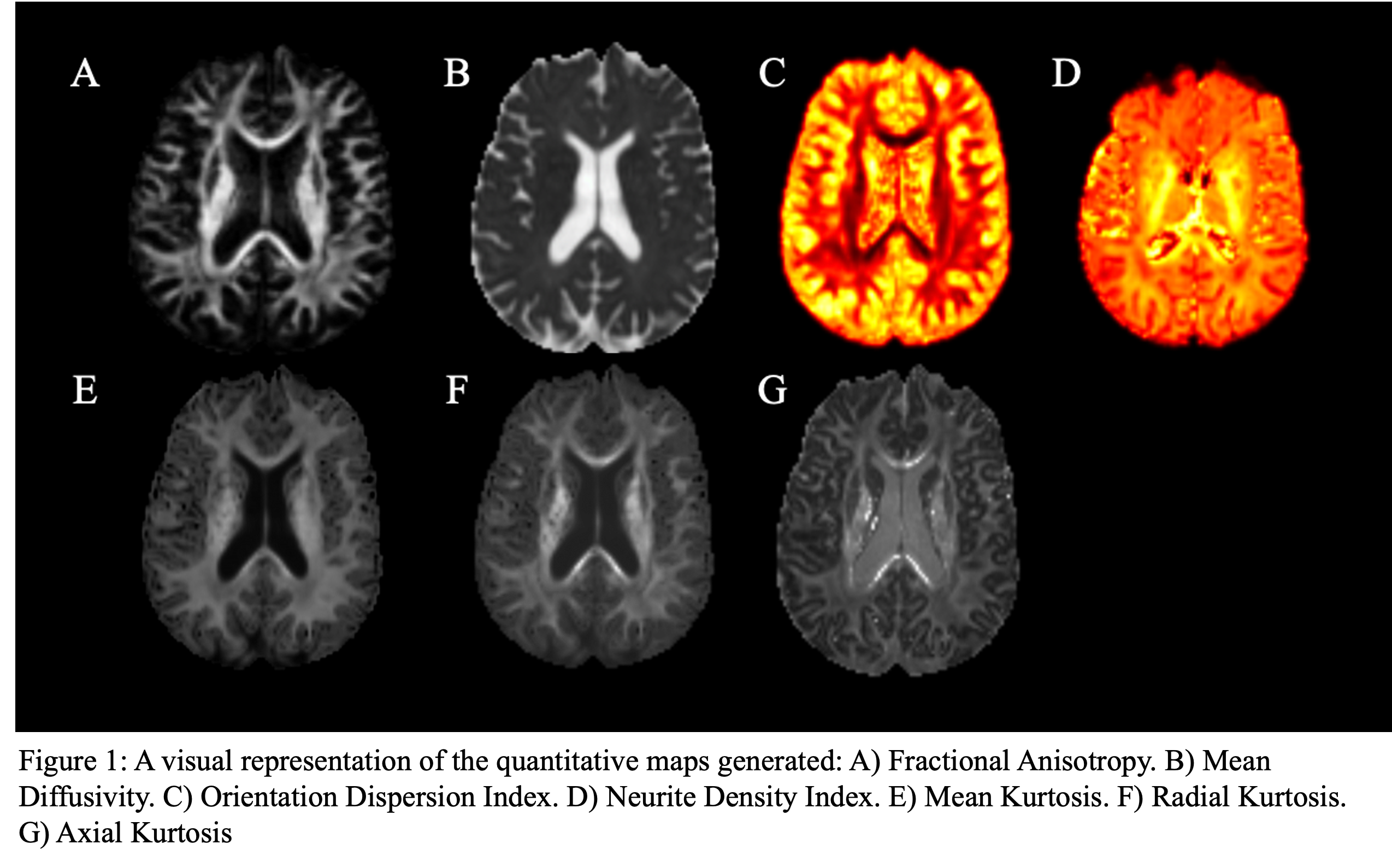
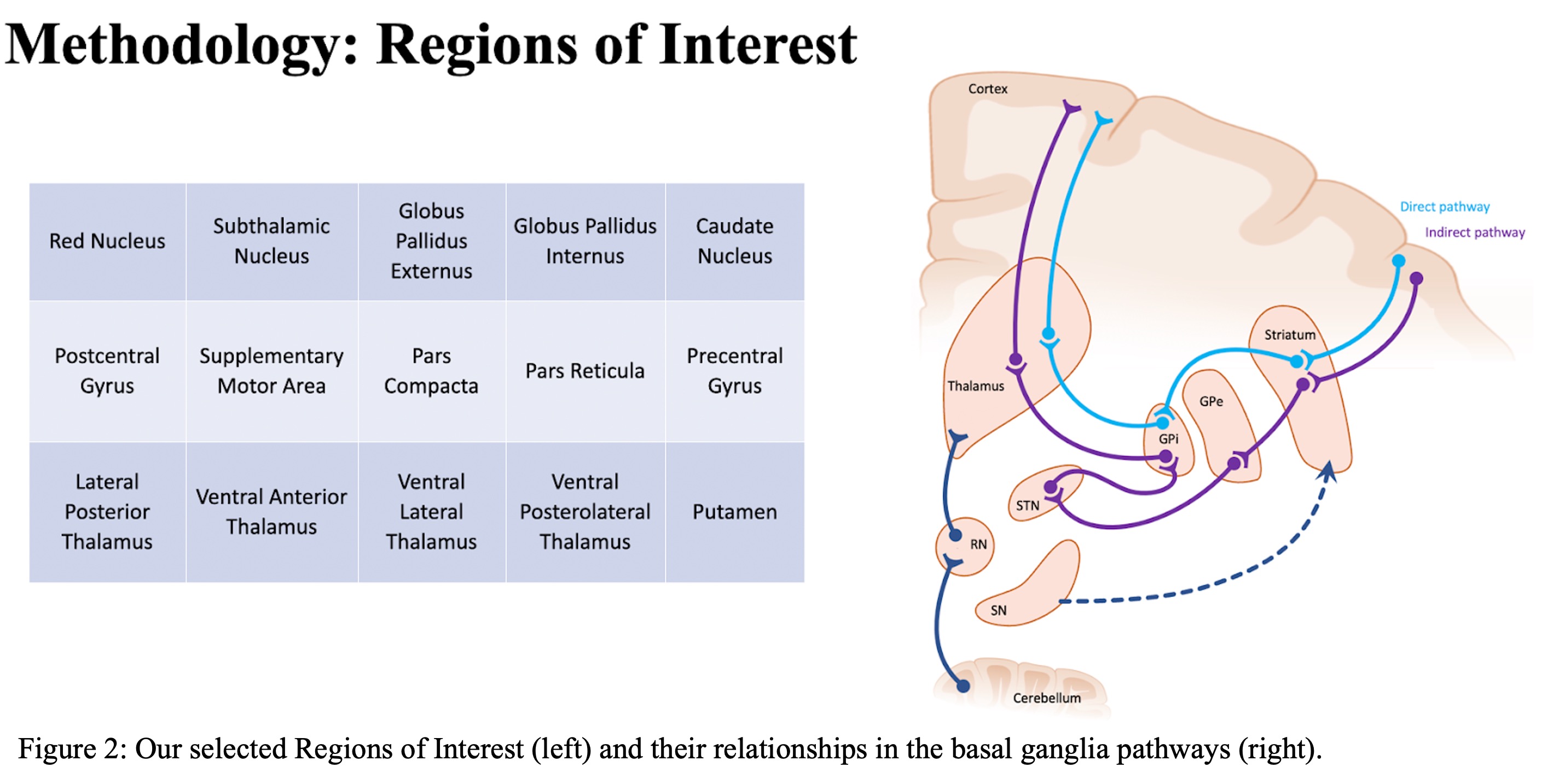
Method: We performed a retrospective study with 43 PD patients (15 females, 28 males, average age = 64). Of these, 11 had NGD, 23 had GD, and 9 had FOG. All subjects underwent 3.0T brain diffusion MRI (dMRI) before DBS surgery. Multiple estimations (NODDI, DKI, and DTI) were applied to the preprocessed data resulting in 7 quantitative maps (Figure 1). These maps included: (1) fractional anisotropy (FA) and mean diffusivity (MD) derived from DTI; orientation dispersion index (ODI) and neurite dispersion index (NDI) maps computed from NODDI, and DKI led to axial kurtosis (AK), mean kurtosis (MK), and radial kurtosis (RK). There were 30 regions-of-interest (ROIs) extracted from an AAL3 and basal ganglia atlas; ROIs all pertained to basal ganglia signaling pathways (Figure 2). ROIs were registered to the patient’s diffusion space, and then the mean values of the aforementioned quantitative maps were calculated for each ROI. Finally, the quantitative data and gaits were analyzed via a Pairwise linear regression model using age, sex, and preoperative UPDRS III scores as confounding variables.
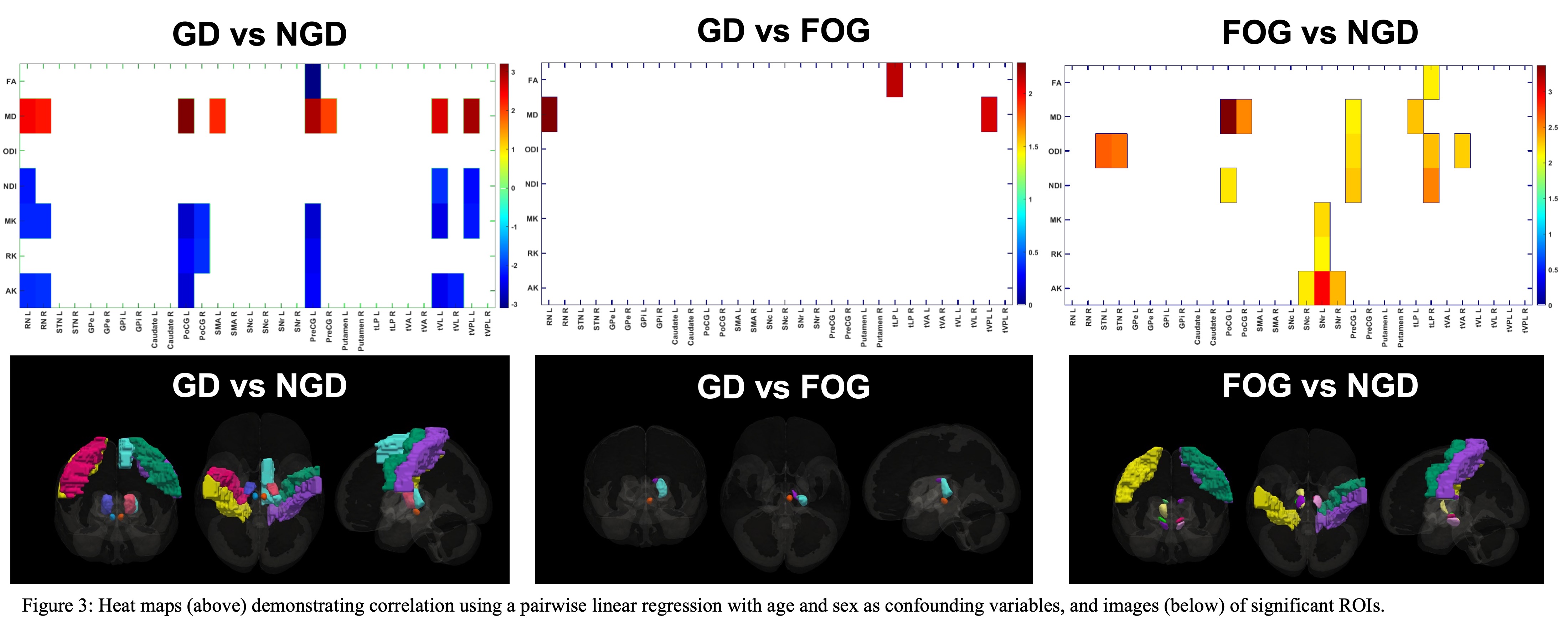
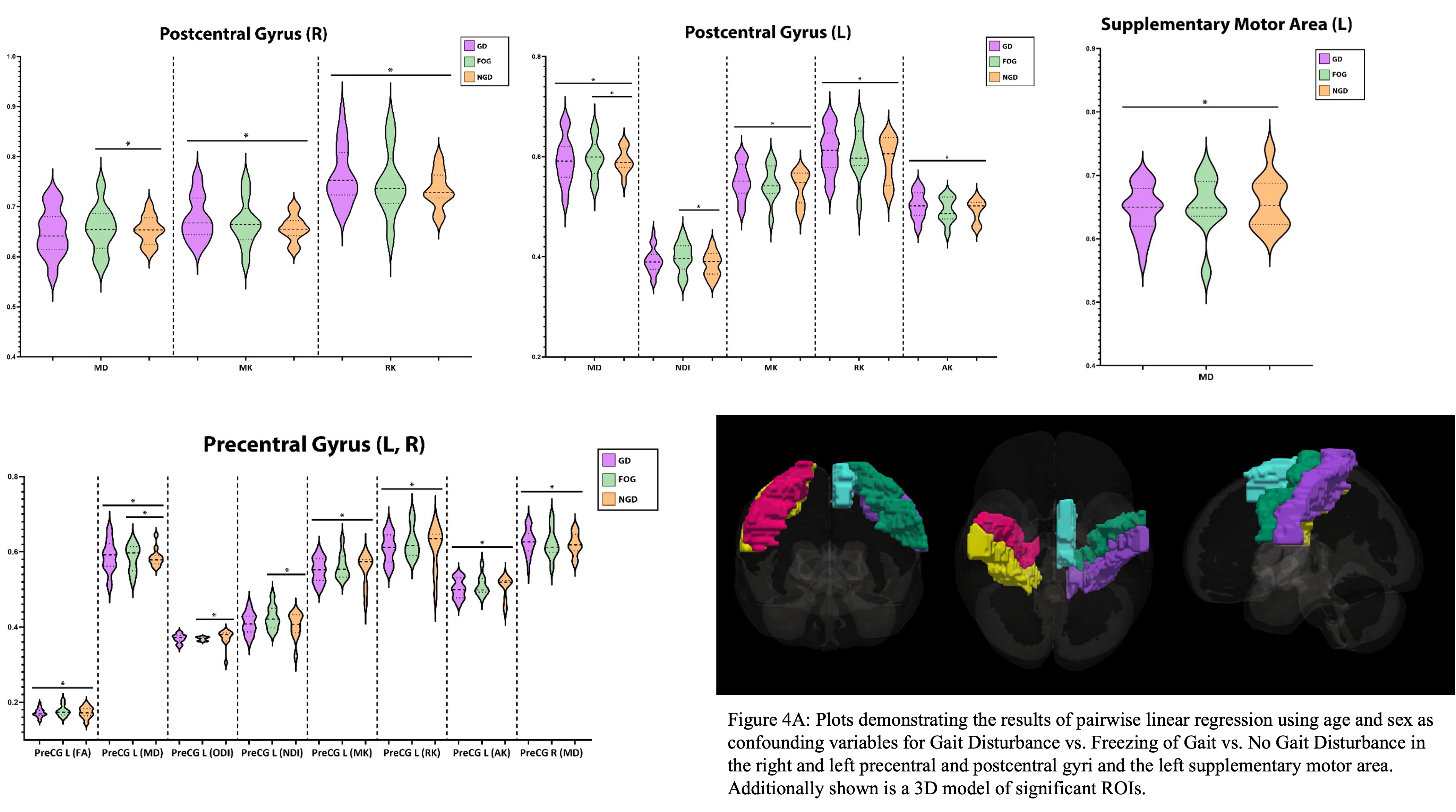
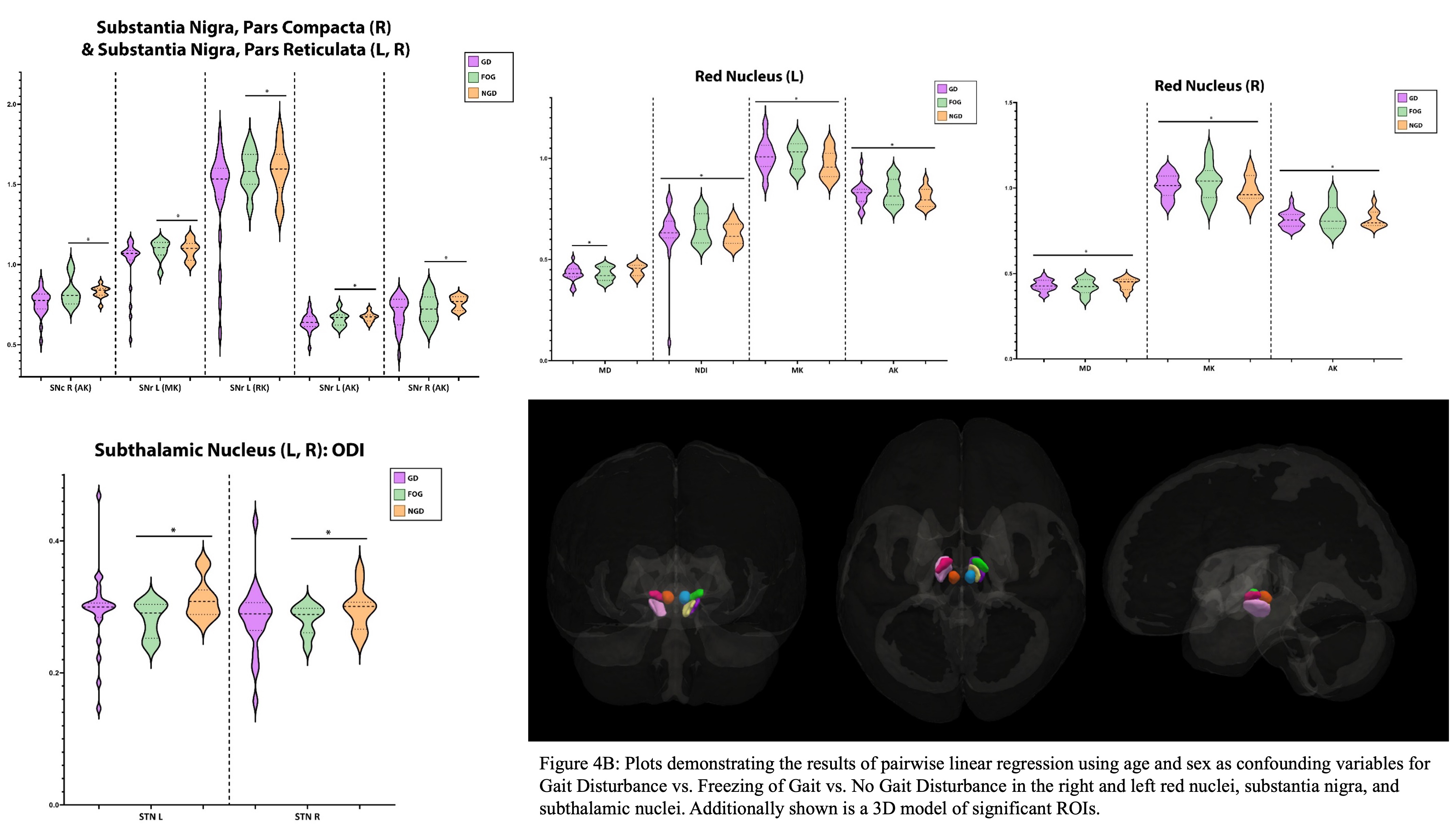
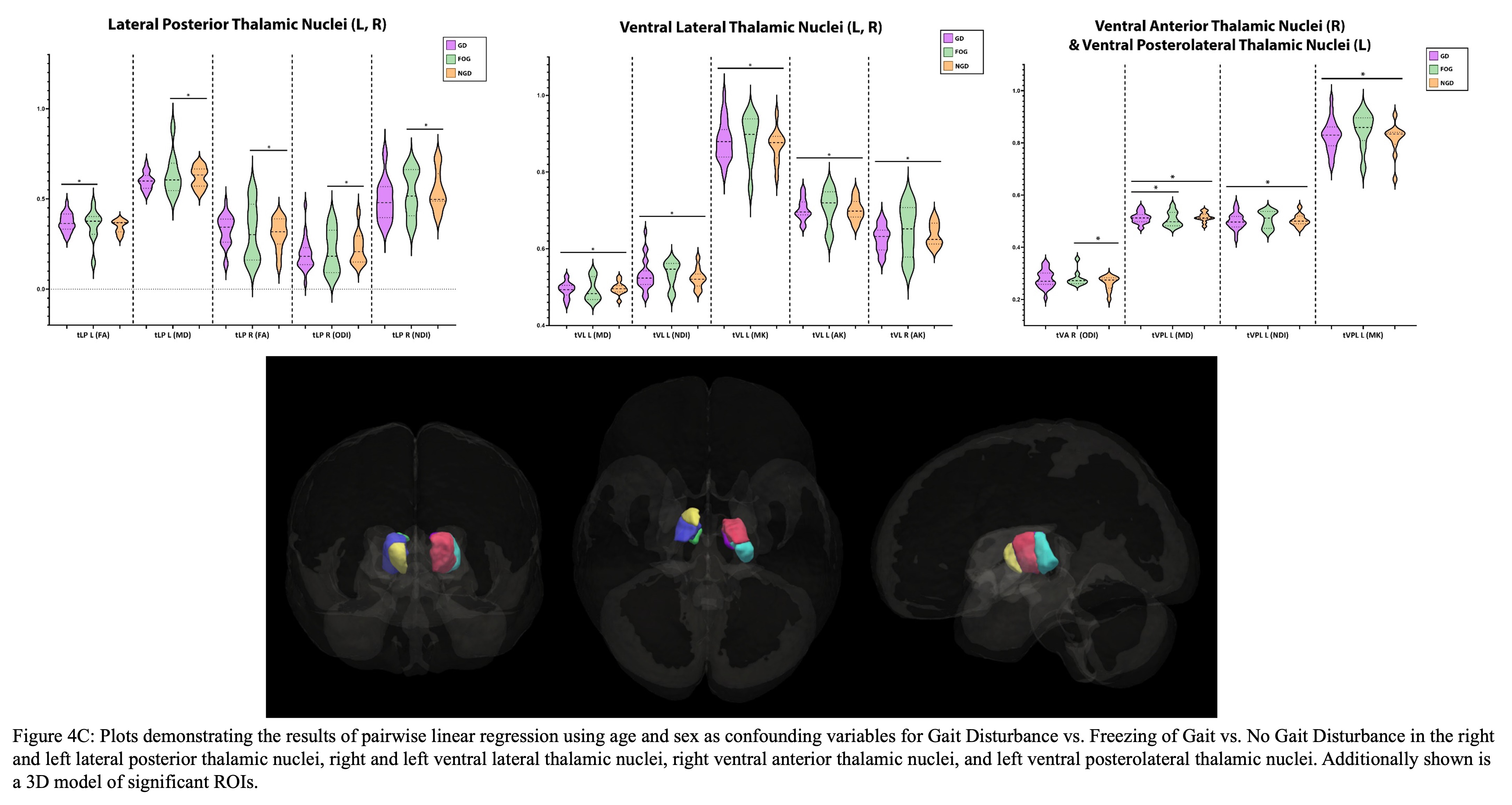
Results: The ROIs with the greatest significance between GD vs. FOG were the left RN, left tLP, and right tVPL (Figure 3A). Between NGD and GD, the left PoCG, right tVPL, and left PreCG showed the most significance (Figure 3B). The comparison of NGD vs. FOG demonstrated the greatest significance in the left PoCG and left SNr (Figure 3C).
Conclusion: Based on these results, quantitative mapping could non-invasively assess the risk and prognosis of gait issues in PD. We hope to create a predictive model to identify a patient’s likelihood of developing GD or FOG.
To cite this abstract in AMA style:
I. Shelley, I. Ailes, M. Syed, K. Shivok, J. Miao, L. Krisa, F. Mohamed, R. Sergott, T. Liang, A. Sharan, M. Kogan, C. Wu, M. Alizadeh, O. Shoraka. Assessing Gait Disturbances in Parkinson’s Disease Using Multi-Spectral Diffusion Weighted Imaging [abstract]. Mov Disord. 2023; 38 (suppl 1). https://www.mdsabstracts.org/abstract/assessing-gait-disturbances-in-parkinsons-disease-using-multi-spectral-diffusion-weighted-imaging/. Accessed December 16, 2025.« Back to 2023 International Congress
MDS Abstracts - https://www.mdsabstracts.org/abstract/assessing-gait-disturbances-in-parkinsons-disease-using-multi-spectral-diffusion-weighted-imaging/

