Category: Surgical Therapy: Parkinson's Disease
Objective: To evaluate distinct anatomical substrates modulating motor subcomponents of bradykinesia following Subthalamic Nucleus (STN) Deep Brain Stimulation (DBS) in Parkinson’s Disease (PD).
Background: PD bradykinesia is rated by a composite of slowed frequency, decrement in amplitude, and loss of rhythm. Understanding whether its components of frequency and amplitude segregate anatomically within basal ganglia structures could facilitate tailored DBS programming to target patient-specific symptoms. In this preliminary report, we investigated the anatomical neural substrates associated with these two components of bradykinesia when STN-DBS was applied.
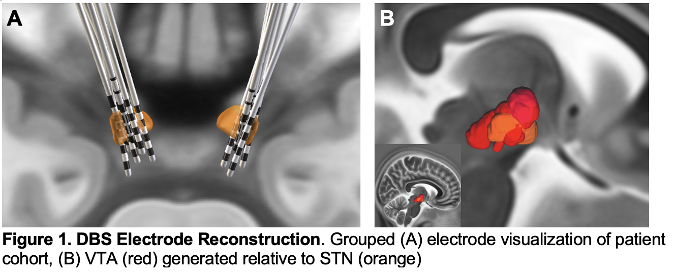
Method: 11 PD patients with STN-DBS were recruited. Using a motion sensor, amplitude and frequency of motion were detected across Finger Tapping (FT), Grasping (GP), Palm Rotation (PR) tasks. In parallel, a trained rater evaluated MDS-UPDRS-III score. Measurements were made in both DBS-ON and DBS-OFF conditions in a randomized order blinded to the rater. The percent change in MDS-UPDRS-III, amplitude, and frequency post-DBS was evaluated. After reconstructing DBS electrodes, the Volume of Tissue Activation (VTA) of each subject was created [figure1]. The change in motor measures was projected to VTAs to create mean effect images to identify distinct anatomical substrates modulating motor subcomponents when stimulated.
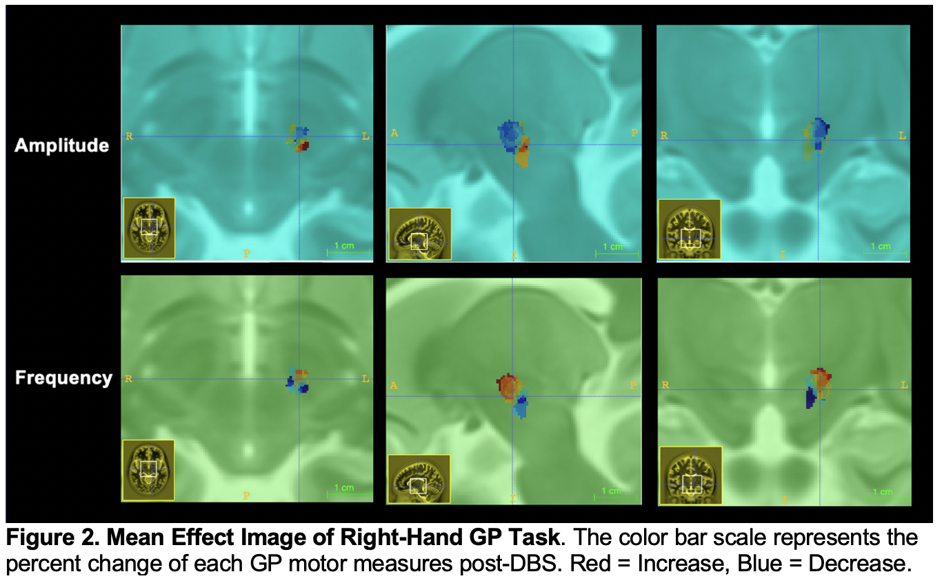
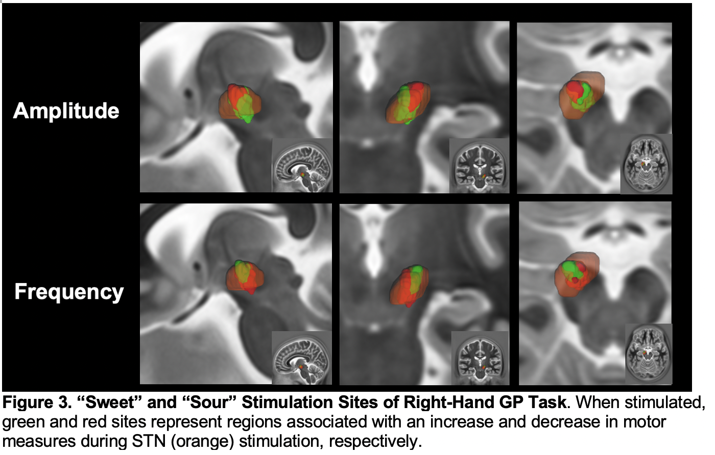
Results: The change in frequency was associated with change in MDS-UPDRS-III right hand GP (β = 2.95, p < 0.05) and PR (β = -1.22, p < 0.05) task post-DBS. The change in frequency was negatively correlated with amplitude for right-hand GP task (r = -0.22, p = 0.009). In the mean effect image, the change in amplitude and frequency was represented as a continuous gradient distributed across the dorsolateral-ventromedial STN [figure2]. Once thresholding the mean effect image to positive and negative percent change in GP task, a clear spatial dichotomy within measure was observed [figure3].
Conclusion: DBS-associated improvement of MDS-UPDRS III was more strongly associated with a change in frequency than amplitude of rapid movement tasks. Degree of frequency improvement was topographically represented in the STN and was separate from that of amplitude improvement. Furthermore, stimulation sites associated with increased frequency were spatially overlapping with that with decreased amplitude.
To cite this abstract in AMA style:
MJ. Kim, Y. Shi, Y. Salimpour, WS. Anderson, K. Mills. Anatomical Substrates for Motor Subcomponents of Bradykinesia in Parkinson’s Disease after Subthalamic Nucleus Deep Brain Simulation [abstract]. Mov Disord. 2022; 37 (suppl 2). https://www.mdsabstracts.org/abstract/anatomical-substrates-for-motor-subcomponents-of-bradykinesia-in-parkinsons-disease-after-subthalamic-nucleus-deep-brain-simulation/. Accessed January 17, 2026.« Back to 2022 International Congress
MDS Abstracts - https://www.mdsabstracts.org/abstract/anatomical-substrates-for-motor-subcomponents-of-bradykinesia-in-parkinsons-disease-after-subthalamic-nucleus-deep-brain-simulation/


