Category: Parkinsonism, Atypical: PSP, CBD
Objective: In this observational/cross-sectional study, we tested whether the degree and extent of in vivo synaptic loss differed in patients with corticobasal syndrome according to their amyloid status: comparing those with likely Alzheimer’s disease (AD) to non-AD pathology.
Background: The corticobasal syndrome (CBS) is a complex asymmetric movement disorder, with cognitive change. Although commonly associated with the primary tauopathy of corticobasal degeneration, clinicopathological correlation is poor. AD pathology causes about one third of CBS. Synaptic loss is a pathological feature of many neurodegenerative disorders[1,2]; it occurs early in disease and is highly related to cognitive function.
Method: 25 people with a clinical diagnosis of possible/probable CBS, and 32 age-/sex-/education-matched healthy volunteers were recruited from tertiary referral centres, and the National Institute for Health Research ‘Join Dementia Research’ platform, respectively. All participants underwent positron emission tomography with [11C]UCB-J and [18F]AV-1451, structural MRI and neuropsychological testing. Participants with CBS had amyloid imaging with [11C]PiB PET. Regional synaptic density was estimated by [11C]UCB-J non-displaceable binding potential (BPND, with and without partial volume correction), AD-tau pathology with [18F]AV-1451 BPND, and grey matter volume with T1-weighted MRI. Symptom severity was assessed with the PSP and the corticobasal ganglia functional rating scales, and the revised Addenbrooke’s Cognitive Examination. Regional differences in synaptic density, [18F]AV-1451 binding and grey matter volume were calculated using ANOVA with age (and where appropriate intracranial volume) as covariates.
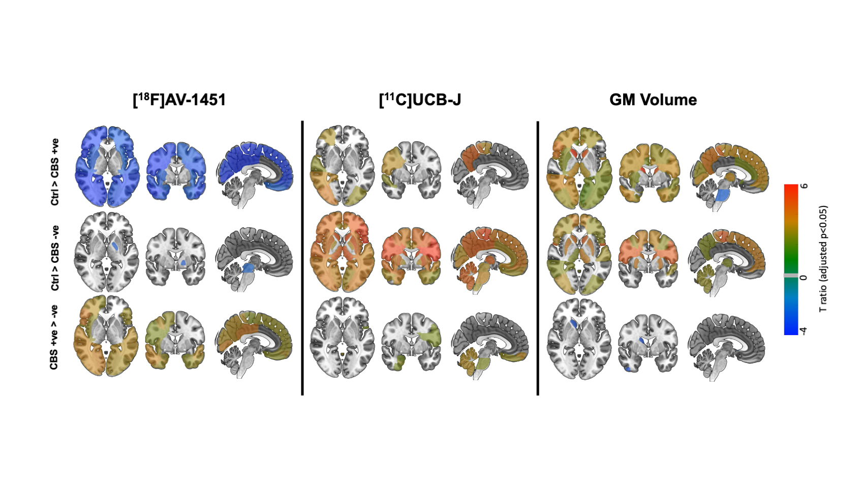
Results: People with CBS had reduced [11C]UCB-J BPND with asymmetry in line with their clinically most affected side. Synaptic loss was more extensive and severe in amyloid-ve patients, with less severe atrophy on MRI[figure1]. Amyloid+ve patients had synaptic loss mainly in posterior cortical regions, in contrast to the more severe anterior and subcortical loss in amyloid-ve patients.
Conclusion: There is severe and asymmetric synaptic loss in CBS. Distinct patterns of synaptic loss, [18F]AV-1451 binding and atrophy, indicate differences in the pathogenic mechanisms based on whether CBS is likely caused by Alzheimer’s disease or corticobasal degeneration. This highlights the need for different therapeutic strategies.
References: [1] Holland N, Jones PS, Savulich G, et al. Synaptic Loss in Primary Tauopathies Revealed by [(11) C]UCB-J Positron Emission Tomography. Mov Disord. 2020;35(10):1834-1842.
[2] Malpetti M., Jones PS, Cope T, Holland N, et al. Synaptic loss in frontotemporal dementia revealed by [(11) C]UCB-J PET. Ann Neurol. 2022;93:142-154.
To cite this abstract in AMA style:
N. Holland, G. Savulich, P. Jones, D. Whiteside, D. Street, M. Naessens, M. Malpetti, Y. Hong, T. Fryer, T. Rittman, E. Mulroy, F. Aigbirhio, K. Bhatia, J. O'Brien, J. Rowe. Amyloid status differentiates synaptic loss and atrophy in Corticobasal Syndrome. [abstract]. Mov Disord. 2023; 38 (suppl 1). https://www.mdsabstracts.org/abstract/amyloid-status-differentiates-synaptic-loss-and-atrophy-in-corticobasal-syndrome/. Accessed December 30, 2025.« Back to 2023 International Congress
MDS Abstracts - https://www.mdsabstracts.org/abstract/amyloid-status-differentiates-synaptic-loss-and-atrophy-in-corticobasal-syndrome/

