Category: Parkinson's Disease: Neuroimaging
Objective: Depression is a common non-motor symptom in Parkinson’s disease (PD), while its underlying pathophysiology remains unclear. Previous functional magnetic resonance imaging (fMRI) studies reported a variety of brain areas may be related to depression in PD, however, the results vary greatly among studies.
Background: Depression is a common non-motor symptom in Parkinson’s disease (PD), while its underlying pathophysiology remains unclear. Previous functional magnetic resonance imaging (fMRI) studies reported a variety of brain areas may be related to depression in PD, however, the results vary greatly among studies.
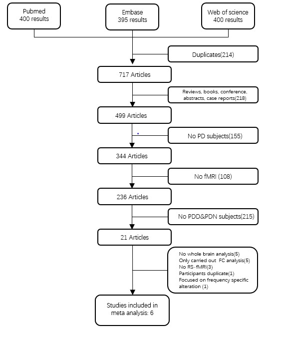
Method: Using signed differential mapping (SDM), we firstly performed a voxel-based meta-analysis in resting-state fMRI (rs-fMRI) studies of depression in PD. Then, ten PD patients with depression (PDD) and 10 PD patients without depression (PDN) performed rs-fMRI after an overnight withdrawal of dopaminergic treatment. Multiscale entropy (MSE) was calculated with region-of-interests (ROIs) based analysis of rs-fMRI in PDD and PDN groups and was then compared within group-level. The ROIs were defined based on the results of our meta-analysis.
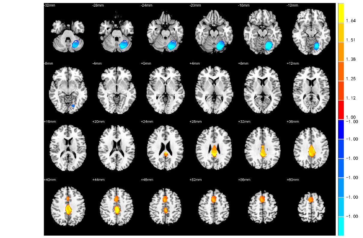
Results: First, the meta-analysis encompassed data from 126 PDD and 153 PDN patients reported in 6 rs-fMRI studies. It yielded neural activity alterations in posterior cingulate gyrus, supplementary motor area (SMA) and cerebellum in PDD. Then, defining these three regions as ROIs, we conducted a ROI-based analysis comparing our newly recruited PDD and PDN groups. We found that compared to PDN, MSE decreased significantly in posterior cingulate gyrus (F=3.747, p=0.002), SMA (F=7.249, p=0.002) and cerebellum (F=0.047, p=0.034) in PDD.
Conclusion: In conclusion, our meta-analysis revealed that specific resting-state brain activity alterations in cingulate gyrus, SMA and cerebellum in PDD. And our ROI-based fMRI analysis provided primary evidence that complexity of neural activity altered in cingulate gyrus, SMA and cerebellum. Quantification of these regional activity changes could serve as a biomarker for PDD and as such may be helpful in unravelling its pathophysiology.
References: Wang, M., et al., Changed Resting-State Brain Signal in Parkinson’s Patients With Mild Depression. Front Neurol, 2020. 11: p. 28. Luo, C., et al., Resting-state fMRI study on drug-naive patients with Parkinson’s disease and with depression. J Neurol Neurosurg Psychiatry, 2014. 85(6): p. 675-83.
To cite this abstract in AMA style:
D. Su, Y. Cheng, J. Zhou, T. Feng. Altered Resting-state Neural Activity in Parkinson’s Disease with Depression: a controlled study based on original meta-analysis results [abstract]. Mov Disord. 2021; 36 (suppl 1). https://www.mdsabstracts.org/abstract/altered-resting-state-neural-activity-in-parkinsons-disease-with-depression-a-controlled-study-based-on-original-meta-analysis-results/. Accessed April 26, 2025.« Back to MDS Virtual Congress 2021
MDS Abstracts - https://www.mdsabstracts.org/abstract/altered-resting-state-neural-activity-in-parkinsons-disease-with-depression-a-controlled-study-based-on-original-meta-analysis-results/


