Session Information
Date: Monday, October 8, 2018
Session Title: Parkinson's Disease: Neuroimaging And Neurophysiology
Session Time: 1:15pm-2:45pm
Location: Hall 3FG
Objective: we investigate the functional connectivity (FC) patterns of the putamen between different subtypes of PD) and healthy controls, and explore its clinical significance.
Background: Impairment of the basal ganglia-thalamo-cortical circuit causes motor symptoms of Parkinson’s disease(PD).
Methods: 16 tremor dominant (TD) PD patients, 23 postural instability and gait difficulty dominant PD patients and 31 healthy controls, were scanned with resting-state functional magnetic resonance imaging. A voxel-wise FC analysis was performed by computing the temporal correlation between the bilateral putamen and the other voxels within the whole brain. Correlation analysis was performed between the FC strength and motor symptoms.
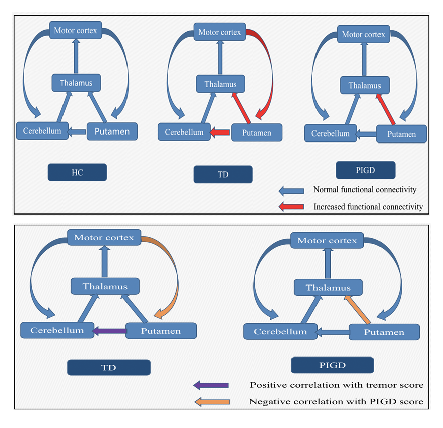
Results: Compared with PIGD dominant group, TD dominant group showed increased FC between bilateral putamen and right cerebellum lobules VI, cerebelum crus I. While compared with healthy controls, tremor dominant PD patients showed increased FC between left putamen and bilateral cerebellum crus I, right cerebelum lobules VI, right thalamus, left paracentral lobule, right inferior occipital lobule, cerebellar vermis, fusiform gyrus, left supplementary motor area. Increased FC between right putamen and right cerebellum crus I, right cerebellum lobules VI, right thalamus, bilateral paracentral lobule, right inferior occipital lobule, right inferior temporal gyrus, bilateral supplementary motor area and left sensorimotor cortex were also shown in TD patients, . PIGD dominant group showed increased FC between right putamen and right thalamus compared with healthy controls. The FC strength between the left putamen and right cerebelum lobules VI showed positive correlation with tremor scores in TD dominant group. The FC strength between right putamen and left sensorimotor cortex showed negative correlation with PIGD scores. While in PIGD dominant group, the FC strength between left putamen and right thalamus showed negative correlation with TD scores.
Conclusions: The altered connectivity of basal ganglia-cortical circuit in PD patients was related to PIGD symptoms, Compared to PIGD patients, motor and cognitive impairment declined slowly in TD patients, Which may be related to the increased functional connectivity between basal ganglia and cerebellum.
References: [1] O’Reilly J X, Beckmann C F, Tomassini V, Ramnani N, Johansen-Berg H. Distinct and overlapping functional zones in the cerebellum defined by resting state functional connectivity[J]. Cereb Cortex,2010,20(4):953-965. [2] Bostan A C, Dum R P, Strick P L. Cerebellar networks with the cerebral cortex and basal ganglia[J]. Trends Cogn Sci,2013,17(5):241-254.
To cite this abstract in AMA style:
B. Shen, L. Zhang. Altered putamen and cerebellum connectivity between different subtypes of Parkinson’s disease [abstract]. Mov Disord. 2018; 33 (suppl 2). https://www.mdsabstracts.org/abstract/altered-putamen-and-cerebellum-connectivity-between-different-subtypes-of-parkinsons-disease/. Accessed April 26, 2025.« Back to 2018 International Congress
MDS Abstracts - https://www.mdsabstracts.org/abstract/altered-putamen-and-cerebellum-connectivity-between-different-subtypes-of-parkinsons-disease/

