Category: Parkinson's Disease: Neuroimaging
Objective: Parkinson’s disease (PD) is frequently accompanied by visual disturbances (VD) and visual hallucinations (VH). This study aimed to analyze the alterations of Dynamic Functional Connectivity (dFC) in subjects with PD and concomitant VD and/or VH compared to no visual disorder (ND). We hypothesized that the commonality in the results between VHD vs VD/ND analysis is due to hallucination.
Background: Visual dysfunction represents a notable non-motor symptom of Parkinson’s disease, yet its understanding remains inadequate. There is scarce research exploring the impact of vision from the brain dynamic networks prospective.
Method: 18 PD subjects with VD (n=8), and concomitant VH and/or VD (VHD; n=8), and 21 PD subjects with ND were enrolled. Resting-state fMRI was performed, and group ICA analysis was executed using GIFT toolbox. 45 components were extracted based on CONN atlas. The Auditory network (AUD; Mid-Superior temporal cortex) and Basal Ganglia (BG) were added separately. dFC analysis was performed, using the sliding window method, followed by k-means clustering (window size = 30, TRs=44, sliding in 1 TR=2ms step). A two-sample t-test was performed to compare the dFC between groups (P<0.05). The number of transitions between states was calculated and compared between groups using two-way ANOVA. (P<0.05)
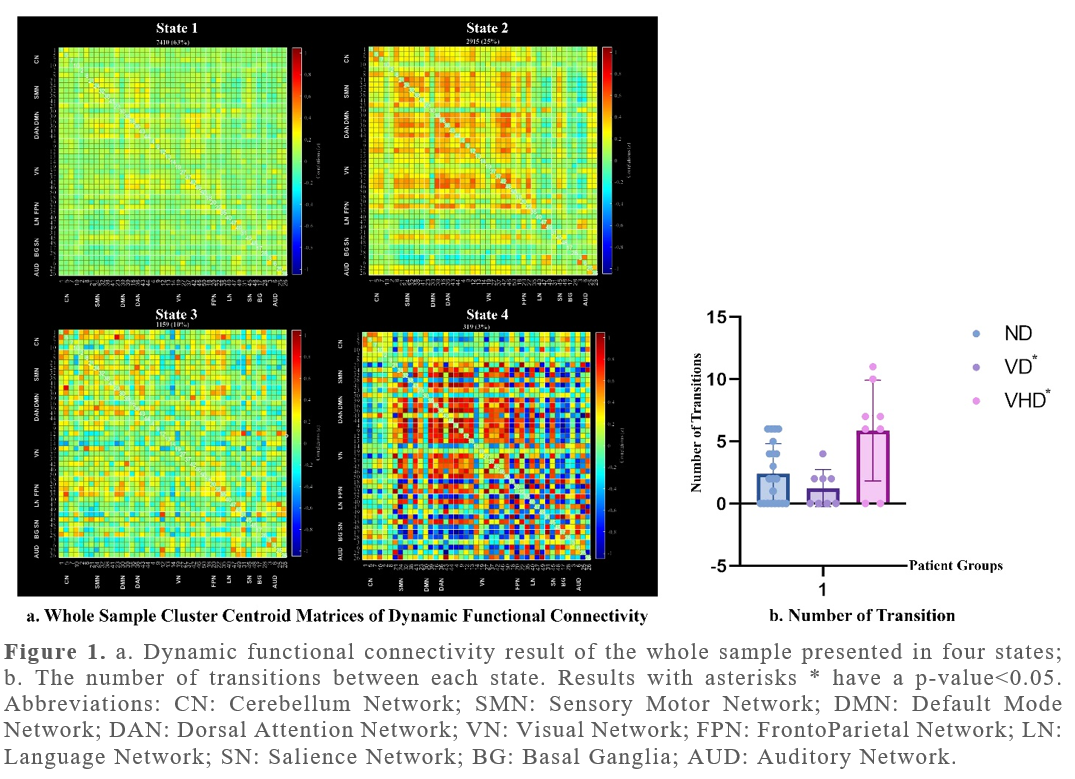
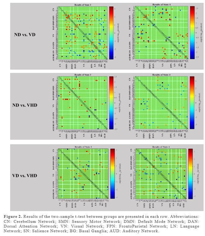
Results: 4 reoccurring FC “States” were identified for the whole sample. The number of transitions between states was significantly higher in the VHD group (results in Figure 1 with asterisks*). dFC differences between each pair of groups in states 1 and 4 were statistically significant. These results are shown in Figure 2.
Conclusion: Hallucination seems to decrease FC both within Visual Network (VN) and between VN and Sensory Motor Network (SMN), FrontoParietal Network (FPN) in state 1, and Language Network (LN) in state 4. Increased FC was shown between Salience Network (SN) and Auditory Network (AUD) in state 1, and between VN and SMN, AUD and LN, and within VN in state 4. Understanding the nature of these alterations can lead to a better insight into the pathophysiology of visual hallucinations.
Figure 1.
Figure2. Results between 3 groups.
To cite this abstract in AMA style:
MA. Alizadeh, SN. Naghizadehkashani, OS. Shoraka, KS. Shivok, IF. Fayed, KH. Hines, AA. Atik, CM. Matias, RS. Sergott, TWL. Liang, CW. Wu. Altered Dynamic Functional Connectivity in Parkinson’s Disease Individuals with Visual Hallucinations and Dysfunction [abstract]. Mov Disord. 2024; 39 (suppl 1). https://www.mdsabstracts.org/abstract/altered-dynamic-functional-connectivity-in-parkinsons-disease-individuals-with-visual-hallucinations-and-dysfunction/. Accessed April 19, 2025.« Back to 2024 International Congress
MDS Abstracts - https://www.mdsabstracts.org/abstract/altered-dynamic-functional-connectivity-in-parkinsons-disease-individuals-with-visual-hallucinations-and-dysfunction/


