Category: Parkinson's Disease: Neuroimaging
Objective: To investigate patterns of altered brain energetics in Parkinson’s disease (PD).
Background: Alterations in mitochondrial function, related to brain energetics, may play an important role in PD initiation/progression. Brain energetics can be quantified via cerebral metabolic rates of oxygen (CMRO2) and glucose (CMRglu) [1]; a lower oxygen-to-glucose index (OGI [2]) can be taken as an index of aerobic glycolysis. OGI varies in the healthy brain, but alterations beyond these may indicate mitochondrial dysfunction or nonneuronal pathology. PD-related metabolism alterations have been robustly identified [3], but OGI alterations have not been fully investigated.
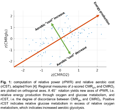
Method: CMRglu and CMRO2 were obtained from 14 PD subjects (disease duration: 6.3 ± 3.7 years) and 10 age-matched healthy controls (HC) during a single scan on the GE SIGNA PET/MR. We decomposed these data into two additional measures: relative power (rPWR, a measure of relative energy production) and relative aerobic cost (rCST, discordance between CMRglu and CMRO2; higher rCST means increased aerobic glycolysis) [Fig. 1]. Scaled Subprofile Modelling Principal Component Analysis (SSM-PCA) was used to identify PD-related regional patterns in CMRglu, CMRO2, rPWR, and rCST. OGI was then computed in affected regions identified through these patterns.
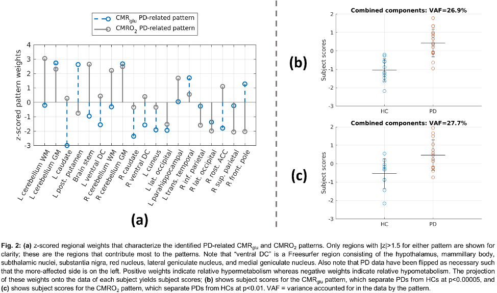
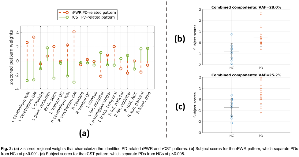
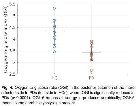
Results: In addition to the well-known regional CMRglu PD-related pattern, we also identify a partially overlapping PD-related CMRO2 pattern strongly separating PDs and HCs [Fig. 2]. PD-related patterns were also identified for rPWR and rCST [Fig. 3]. rPWR is elevated in PDs compared to HCs in cerebellum and lowered in occipitoparietal regions; rCST is elevated in PDs compared to HCs in posterior putamen (more affected side) and lower in cerebellum and brain stem, indicating relatively increased and decreased aerobic glycolysis, respectively. In terms of absolute values, OGI was only significantly different (lowered in PD) in posterior putamen [Fig. 4].
Conclusion: Pattern analysis reveals CMRglu is relatively increased without a corresponding relative increase in CMRO2 in the more affected posterior putamen in PDs, indicating increased aerobic glycolysis which may suggest mitochondrial dysfunction. Conversely, in bilateral cerebellum and brain stem relative increases in CMRO2 exceed relative increases in CMRglu, suggesting high energy production in these regions without relying on aerobic glycolysis.
References: [1] Powers et al. (2008) J Cereb Blood Flow Metab, vol. 28, no. 10, pp. 1754–1760.
[2] Vaishnavi et al. (2010). Proc Natl Acad Sci U S A, vol. 107, no. 41, pp. 17757–17762.
[3] Ma et al. (2010). J Cereb Blood Flow Metab, vol. 30, no. 3, pp. 505–509
[4] E. Shokri-Kojori et al. (2019). Nat Commun, vol. 10, pp. 690, Feb 2019.
To cite this abstract in AMA style:
C. Bevington, J-C. Cheng, R. Jayde-Williams, B. Pike, J. Mckenzie, A. Stoessl, V. Sossi. Altered brain energetics in Parkinson’s Disease: a multimodal neuroimaging study [abstract]. Mov Disord. 2023; 38 (suppl 1). https://www.mdsabstracts.org/abstract/altered-brain-energetics-in-parkinsons-disease-a-multimodal-neuroimaging-study/. Accessed April 26, 2025.« Back to 2023 International Congress
MDS Abstracts - https://www.mdsabstracts.org/abstract/altered-brain-energetics-in-parkinsons-disease-a-multimodal-neuroimaging-study/


