Category: Parkinson's Disease: Neuroimaging
Objective: To explore the potential role of the right olfactory orbitofrontal cortex in olfactory dysfunction (OD) in Parkinson’s Disease (PD).
Background: The orbitofrontal cortex (OFC) is one of the main olfactory projections from the primary olfactory cortex1. The olfactory OFC (Oolf) has been associated with conscious olfactory perception and odor discrimination2. Structural and functional imaging studies have revealed disruptions in the right OFC in PD patients with OD3. However, graph analysis has not been used to investigate the properties of this area.
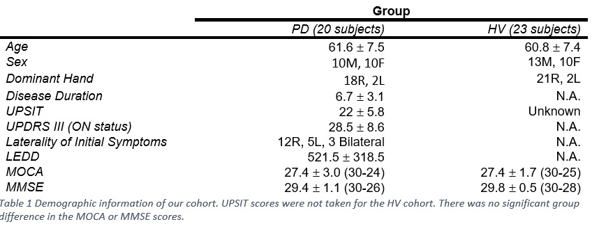
Method: We collected structural MR images and 6-min eye closed rs-fMRI from 30 PD subjects (PDs) in the off-medication state and 24 age/sex matched healthy volunteers (HVs) in a 3T GE MR750 scanner. We administered the University of Pennsylvania Smell Identification Test (UPSIT) to measure olfactory function. (Table 1)
MRI data was preprocessed in AFNI.4 The olfactory network (OlfN) in HVs and PDs was constructed using the human olfactory network template published by TC Arnold5. We generated our network with 19 nodes. The Python version of the Brain Connectivity Toolbox6 was used to calculate graph metrics and also employed proportional thresholding to remove weak connections. To identify specific nodes and edges which differ between graphs, as well as nodal properties, we used the Network Based Statistics (NBS)7. We used the FDR correction to control for false positives.
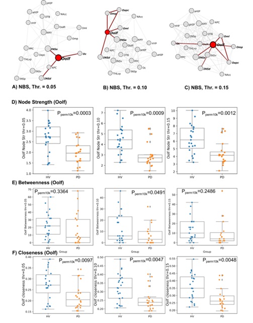
Results: We created our own composite score (hub score) to rank nodes in the network based on the nodal properties. The right Oolf had a low hub score and had the most group difference at all thresholds. The right Oolf also had lower node strength, lower betweenness centrality, and lower closeness in PD compared to HV (Figure 1). NBS failed to identify any left Oolf edges as significantly impacted between groups.
Conclusion: The right Oolf has been associated with conscious olfactory perception and odor discrimination in normal subjects. Alterations to this area, might contribute to olfactory dysfunction in PD.
References: 1. Gottfried, J. A. (2010). Central mechanisms of odour object perception. Nature Reviews. Neuroscience, 11(9), 628. https://doi.org/10.1038/NRN2883
2. Li, W., Lopez, L., Osher, J., Howard, J. D., Parrish, T. B., & Gottfried, J. A. (2010). Right Orbitofrontal Cortex Mediates Conscious Olfactory Perception. Psychological Science, 21(10), 1454. https://doi.org/10.1177/0956797610382121
3. Georgiopoulos, C. (2019). Imaging Studies of Olfaction [Dissertation, Linköping University]. www.liu.se
4. Cox, R. W. (1996). AFNI: Software for Analysis and Visualization of Functional Magnetic Resonance Neuroimages. Computers and Biomedical Research, 29(3), 162–173.
5. Arnold, T. C., You, Y., Ding, M., Zuo, X. N., de Araujo, I., & Li, W. (2020). Functional connectome analyses reveal the human olfactory network organization. ENeuro, 7(4), 1–14. https://doi.org/10.1523/ENEURO.0551-19.2020
6. Rubinov, M., & Sporns, O. (2010). Complex network measures of brain connectivity: Uses and interpretations. NeuroImage, 52(3), 1059–1069. https://doi.org/10.1016/J.NEUROIMAGE.2009.10.003
7. Zalesky, A., Fornito, A., & Bullmore, E. T. (2010). Network-based statistic: Identifying differences in brain networks. NeuroImage, 53(4), 1197–1207. https://doi.org/10.1016/J.NEUROIMAGE.2010.06.041
To cite this abstract in AMA style:
L. Pesantez Pacheco, M. Saquib, D. Ehrlich, M. Hallett, S. Horovitz. Alterations of the Right Olfactory Orbitofrontal Cortex and olfactory impairment in Parkinson’s Disease [abstract]. Mov Disord. 2023; 38 (suppl 1). https://www.mdsabstracts.org/abstract/alterations-of-the-right-olfactory-orbitofrontal-cortex-and-olfactory-impairment-in-parkinsons-disease/. Accessed February 1, 2026.« Back to 2023 International Congress
MDS Abstracts - https://www.mdsabstracts.org/abstract/alterations-of-the-right-olfactory-orbitofrontal-cortex-and-olfactory-impairment-in-parkinsons-disease/


