Category: Choreas (Non-Huntington's Disease)
Objective:
To report case of generalised chorea due to aceruloplasminemia caused by novel mutation in the CP gene
Background: Aceruloplasminemia is a type of Neurodegeneration with Brain Iron Accumulation. It is an autosomal recessive disorder.[1] Impairment ceruloplasmin activity leads to Pathological iron retention and iron-mediated oxidative damage.
Method: We report case of an elderly woman presenting with progressive generalized chorea and cognitive dysfunction clinically mimicking Huntington’s disease. Specific investigations and neuroimaging raised a suspicion of Aceruloplasminemia which was confirmed by genetic testing.
Results: 59 years old woman presented with generalised and eye brow raising chorea with cognitive impairment over 2 years and oromandibular dyskinesias since 6 months. Left hand dystonia and left striatal toe was evident while walking. Cognitive testing revealed Mini Mental Status Examination score of 23/30 and Frontal Assessment Battery score of 11/18 suggestive of frontal-subcortical dysfunction. She had diabetes for 10 years.
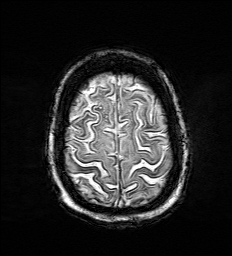
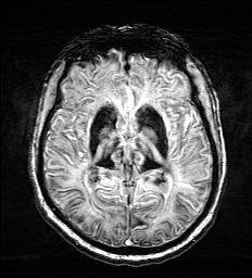
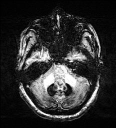
MRI Brain showed T2 hypointensity, SWI blooming in bilateral basal ganglia, substantia nigra, thalamus, dentate nucleus and pencil lining of cortex without any cavitations (figure 1-3). CT brain was normal thus suggestive of iron deposition. Her routine laboratory testing were normal. Serum ceruloplasmin was absent, serum ferritin was high 698ng/ml, low serum iron 24mcg/dl and low transferrin saturation. Peripheral smear for acanthocytes was negative with normal CAG repeats which ruled out other causes of chorea.
Homozygus missense mutation in exon 9 of CP gene (chr3:g.148916189A>G) that results in amino acid substitution of arginine for cysteine at codon 560 (p.cys560Arg) was detected. This variant has not been previously detected hence parents’ variant sequencing is planned.
Clinical presentation of generalised chorea dystonia, absent serum ceruloplasmin, high ferritin with low serum iron, association with diabetes and characteristic MRI brain finding of iron deposition with mutation in CP gene confirms aceruloplasminemia.[2]
Conclusion:
We report a patient with Aceruloplasminemia. Unique features in this case were presentation with generalised chorea with frontal dysfunction mimicking HD phenocopy.[3] This case adds to differentials of HD- Look – Alike. Imaging showed distinctive pencil lining of cortex.[4] Genetic confirmation with mutation in CP gene was obtained.
References: 1. Marchi G, Busti F, Lira Zidanes A, Castagna A and Girelli D (2019) Aceruloplasminemia: A Severe Neurodegenerative Disorder Deserving an Early Diagnosis. Front. Neurosci. 13:325. 2. Pelucchi, S., Mariani, R., Ravasi, G., Pelloni, I., Marano, M., Tremolizzo, L., et al. (2018). Phenotypic heterogeneity in seven Italian cases of aceruloplasminemia. Parkinsonism Relat. Disord. 51, 36–42. 3.Batla A, Adams ME, Erro R, et al. Cortical pencil lining in neuroferritinopathy: a diagnostic clue. Neurology. 2015;84(17):1816–1818. 4. Schneider SA, Bird T. Huntington’s Disease, Huntington’s Disease Look-Alikes, and Benign Hereditary Chorea: What’s New?. Mov Disord Clin Pract. 2016;3(4):342–354. Published 2016 Jan 27.
To cite this abstract in AMA style:
S. Patil, M. Bhatt, A. Aggarwal. Aceruloplasminemia Presenting as Huntington’s Disease –Look –Alike with Unique MRI Features [abstract]. Mov Disord. 2020; 35 (suppl 1). https://www.mdsabstracts.org/abstract/aceruloplasminemia-presenting-as-huntingtons-disease-look-alike-with-unique-mri-features/. Accessed April 19, 2025.« Back to MDS Virtual Congress 2020
MDS Abstracts - https://www.mdsabstracts.org/abstract/aceruloplasminemia-presenting-as-huntingtons-disease-look-alike-with-unique-mri-features/


