Category: Neuroimaging (Non-PD)
Objective: Reviewing typical neuroimaging features of the substantia nigra.
Noting that the absence of nigrosome 1 has also been seen in atypical parkinsonisms.
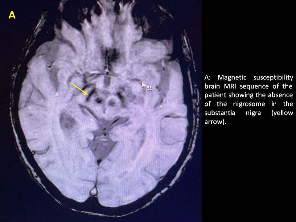
Background: It is known that several neurodegenerative processes are associated with iron accumulation in the CNS,which is equivalent to the loss of dopaminergic neurons.Nigrosomes are small accumulations of dopaminergic neurons located in the substantia nigra of the midbrain.The most relevant is nigrosome 1 which is located in the caudal and central region of the substantia nigra of the midbrain.In PD, it has been described that the absence of nigrosome 1,which is the most affected,is a finding that can be detected in MRI susceptibility sequences and it has been found that its absence has a sensitivity of 100% and a specificity of 84.6% to distinguish PD from healthy people.We describe a patient with clinical symptoms suspicious of CBD versus PSP who presented this radiological sign in the neuroimaging study.
Method: A 75 year old woman with a one-year history of progressive mobility deterioration,with increasingly frequent falls, most of them backwards.She also had changes in facial expression,sometimes with her eyes wide open,as if she were concentrated.She has no tremor.
Neurological examination of the patient was normal except for some apraxia of the gaze;EOMs with supra and infraversion restriction and decreased latency of saccades.Hypomimia with marked decrease in blinking was also seen.She doesn’t raise left upper limb(LUL)spontaneously,when she was asked to raise it she helped herself with the right upper limb(RUL).She cooperated very little although distal segmental strength of all four limbs was 5/5(she didn’t cooperate with proximal LUL).Mild rigidity of LUL although it gives the impression of some left hemineglection.Mild RUL bradykinesia.Gait had short steps,requiring supporting area.Increased dragging of the left foot was also seen.
Results: Clinical findings showed extrapyramidal involvement associated with cortical symptoms,that’s why it was decided to perform a brain MRI to rule out other conditions and findings supporting atypical parkinsonisms.This study showed the absence of nigrosome 1.
Conclusion: Although in most publications the sign of the absence of nigrosome 1 has been associated with PD,this case opens up the possibility that it could be seen in other atypical parkinsonisms and therefore it may not be a differentiating finding between them.
To cite this abstract in AMA style:
L. Caballero Sánchez, D. Cerdán Santacruz, C. Gómez López, J. Berrío Suaza, P. Gil Armada, M. álvarez Eulate, A. Castrillo Sanz, A. Mendoza Rodriguez, M. Rodríguez Sanz, C. Tabernero García. Absence of nigrosome as a finding in a patient with suspected atypical parkinsonism. [abstract]. Mov Disord. 2022; 37 (suppl 2). https://www.mdsabstracts.org/abstract/absence-of-nigrosome-as-a-finding-in-a-patient-with-suspected-atypical-parkinsonism/. Accessed April 18, 2025.« Back to 2022 International Congress
MDS Abstracts - https://www.mdsabstracts.org/abstract/absence-of-nigrosome-as-a-finding-in-a-patient-with-suspected-atypical-parkinsonism/

