Session Information
Date: Thursday, June 8, 2017
Session Title: Dystonia
Session Time: 1:15pm-2:45pm
Location: Exhibit Hall C
Objective: To explore functional connectivity at network level with MEG, pre and post-administration of medications, in generalized and cervical dystonia.
Background: The underlying mechanisms of dystonia remain poorly understood. Mechanisms thought to play a critical role include reduced intracortical inhibition and distorted somatotopic cortical representation. Prior studies showed altered functional connectivity within the sensorimotor, the executive control and the primary visual network.
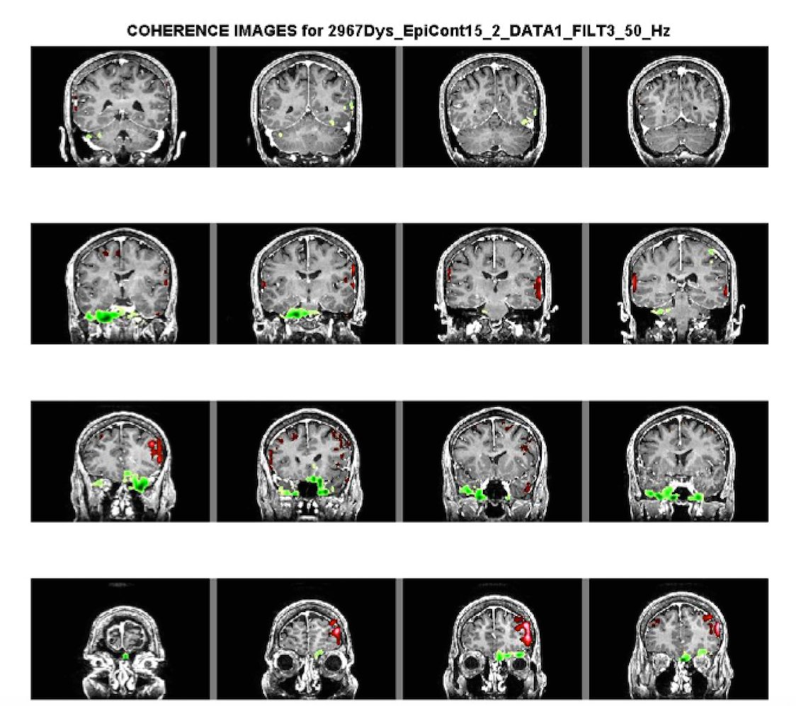
Methods: MEG Data on 5 patients, 3 with cervical and 2 with generalized dystonia (age: 16-70 years) were matched to 4 healthy controls. All patients (3 on Diazepam and baclofen, 1 on Sinemet, 1 on Botulinum toxin) showed clinical benefit with medications. Two 15-minute MEG scans spaced one hour apart in each treatment condition (pre- medication and 1 hour after ingested medication) were obtained. Synchronization of neuronal activity was quantified by calculating coherence source imaging (CSI) between cortical sites from MEG imaged brain activations. A region-of-interest (ROI) tool was used to identify 54 regions in the brain (27 in each hemisphere). Within the frequency band 3-50Hz, a t-test was conducted to assess group difference in average CSI values for each pair of brain regions (N= 1,431). A P value was produced for each region pair. The false discovery rate (FDR) was used to adjust for multiple testing. Only statistically significant coherence values (p<0.05) with a large effect size were considered.
Results: In both pre-treatment and post-treatment groups, the biggest difference in coherence between patients and controls was seen in the occipito-striatal and occipito-parietal regions, with some differences in the fronto-striatal and fronto-occipital regions. On comparing pre- and post-treatment dystonia patients, a decrease in coherence was observed in these regions post treatment.
Conclusions: Our exploratory study using MEG, while limited by a small sample size, showed increased coherence in regions associated with visuospatial and executive function, with a decrease post medication in patients with cervical and generalized dystonia. The latter may be a biomarker for normalization of aberrant connectivity and merits further exploration.
References: 1. Elisevich K, Shukla N, Moran J, et al. An Assessment of MEG Coherence Imaging in the Study of Temporal Lobe Epilepsy. Epilepsia. 2011;52(6):1110-1119. doi:10.1111/j.1528-1167.2011.02990.x. 2. Neychev VK, Gross R, Lehéricy S, Hess EJ, Jinnah HA. The Functional Neuroanatomy of Dystonia. Neurobiology of disease. 2011;42(2):185-201. doi:10.1016/j.nbd.2011.01.026.
To cite this abstract in AMA style:
A. Mahajan, S. Bowyer, A. Zillgitt, P. LeWitt, P. Kaminski, C. Sidiropoulos. A Pilot Multiparametric MEG Study of coherence in generalized and cervical dystonia [abstract]. Mov Disord. 2017; 32 (suppl 2). https://www.mdsabstracts.org/abstract/a-pilot-multiparametric-meg-study-of-coherence-in-generalized-and-cervical-dystonia/. Accessed April 18, 2025.« Back to 2017 International Congress
MDS Abstracts - https://www.mdsabstracts.org/abstract/a-pilot-multiparametric-meg-study-of-coherence-in-generalized-and-cervical-dystonia/

