Session Information
Date: Thursday, June 8, 2017
Session Title: Parkinson's Disease: Neuroimaging And Neurophysiology
Session Time: 1:15pm-2:45pm
Location: Exhibit Hall C
Objective: We model the rate of disease progression over 4 years in subjects with Parkinson’s disease based on neuroimaging features acquired at baseline.
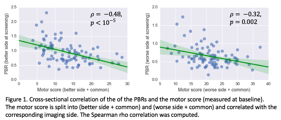
Background: DaTSCAN is a radiotracer with high affinity for the presynaptic dopamine transporter (DAT) in the striatum. The putamen binding ratio (PBR) is the ratio of the tracer uptake between the putamen and the reference region (the occipital cortex). We use PBR data from 95 PD subjects from the PPMI database that have completed scans at baseline (year 0) and at years 1, 2 and 4. At baseline, PBR values were found to be significantly correlated with UPDRS motor score (Figure 1).
[figure1]
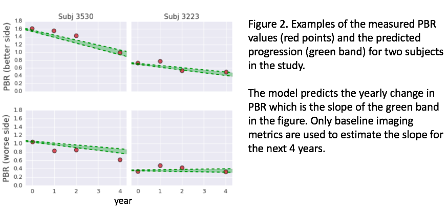
Methods: A hierarchical model is fitted to estimate the rate of progression as estimated by the change in PBR values. Our assumption is that the imaging data would contain relevant and objective information on disease progression as related to dopaminergic deficit. We identify the following image features, all measured at baseline: 1) PBR values for the more and less affected sides, 2) difference between the PBR values of the better and worse sides, and 3) difference between the putamen and caudate binding ratios (PBR – CBR). The model parameters were obtained in a training phase using a subset of the data.
Next, the predicted yearly change of the PBR is estimated for each subject based on the baseline image features only and the estimated model parameters. This yearly change is used to predict the PBR at years 1, 2 and 4 (see Figure 2 for examples). The model is applied separately to the better and to the worse sides of the putamen. Cross-validation (5 folds) was used to estimate the error rate for year-4 PBR predictions.
[figure2]
Results: The median error for year-4 predictions is 18% for the better side and 24% for the worse side (Figure 3) and smaller errors were observed at years 1 and 2. The most important image feature for predicting the progression is the PBR value at baseline. The best predictor is obtained using the better side with the baseline PBR values (p < 0.001) and the PBR difference between the better and worse (p < 0.01). The (PBR – CBR) term is not significant.
[figure3]
Conclusions: A model is developed that predicts disease progression over 4 years as quantified by PBR values with 18% accuracy. In addition to baseline PBR values, this model includes the feature reflecting the side-to-side asymmetry. Such asymmetry is known to be a function of disease manifestation and progression.
To cite this abstract in AMA style:
N. Shenkov, I. Klyuzhin, A. Rahmim, V. Sossi. A neuroimaging-based model for disease progression in Parkinson’s disease [abstract]. Mov Disord. 2017; 32 (suppl 2). https://www.mdsabstracts.org/abstract/a-neuroimaging-based-model-for-disease-progression-in-parkinsons-disease/. Accessed April 16, 2025.« Back to 2017 International Congress
MDS Abstracts - https://www.mdsabstracts.org/abstract/a-neuroimaging-based-model-for-disease-progression-in-parkinsons-disease/


