Category: Parkinson's Disease: Non-Motor Symptoms
Objective: To evaluate corneal sub-basal nerve plexus density in PD compared to age-matched healthy subjects using confocal microscopy
Background: Cornea is the most densely innervated structure (1). One of the objective methods to evaluate the corneal nerve is the implication of confocal microscopy. For the corneal nerve study, the sub-basal nerve plexus is the most common location to analyze due to its dense plexus and the easiest to capture. The nerve density decreases overtime without gender association (2). There were few studies of corneal nerve in PD (3-8). There were consistent results of decreased corneal nerve fiber density (4, 7). They found the correlations between corneal nerve parameters and the motor, autonomic symptoms, and cognitive score (4, 5, 7). However, there were few studies with small sample size.
Method: Ten PD and age-matched healthy controls were recruited into a cross-sectional pilot study. The exclusion criteria were diabetes mellitus, Sjogren syndrome, post-refractive surgery, post-cataract surgery. 75-gram Oral Glucose Tolerant Test (OGTT) was performed to exclude impaired fasting glucose subjects. Demographic data and nerve density were evaluated by a corneal nerve expert ophthalmologist.
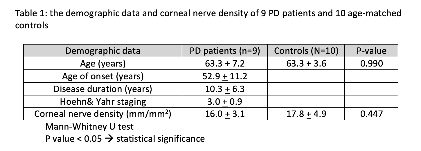
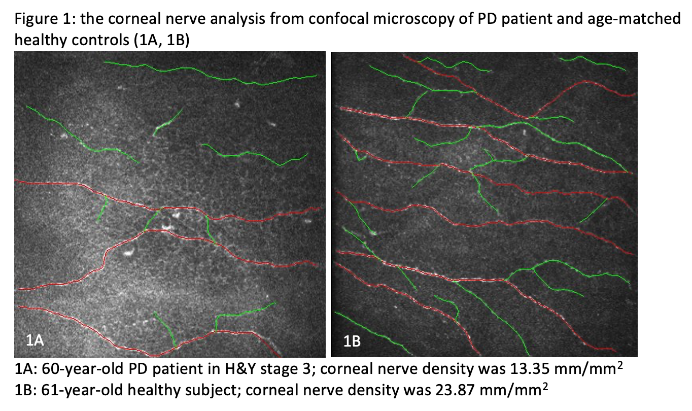
Results: One PD patient was excluded due to the abnormal OGTT test. The mean age of PD and controls were 63.3+7.2 vs 63.3+3.6 years (P=0.990). The mean age of onset, disease duration, UPDRSIII and H&Y staging of PD patients was 52.9+11.2 years, 10.3+6.3 years, 23.2+11.6, and 3.0+0.9, respectively. The mean density in PD patients compared to control group were 16.0+3.1 vs 17.8+4.9 mm/mm2 (P= 0.447). The mean density in H&Y stage 2 was 17.0+2.5 mm/mm2. The mean density in H&Y stage 3 was 16.5+4.4 mm/mm2. The mean density in H&Y stage 4 was 14.5+2.2 mm/mm2. We can see the trend of decreased nerve density along with H&Y grading (one-way ANOVA, P=0.615). A significant correlation between UPDRS part III and nerve density was found (r= -0.683, P= 0.042).
Conclusion: In our pilot study, corneal sub-basal nerve plexus density in PD patients was found to decrease compared to the control subject. There was a trend of decreased corneal nerve density along with PD severity. The corneal parameter could be a potential biomarker to demonstrate the disease severity. A further large sample-size study is necessary to evaluate the correlation with statistical analysis to demonstrate the significant difference.
References: 1. Al-Aqaba MA, Dhillon VK, Mohammed I, Said DG, Dua HS. Corneal nerves in health and disease. Prog Retin Eye Res. 2019;73:100762. 2. Niederer RL, Perumal D, Sherwin T, McGhee CN. Age-related differences in the normal human cornea: a laser scanning in vivo confocal microscopy study. Br J Ophthalmol. 2007;91(9):1165-9. 3. Podgorny PJ, Suchowersky O, Romanchuk KG, Feasby TE. Evidence for small fiber neuropathy in early Parkinson’s disease. Parkinsonism Relat Disord. 2016;28:94-9. 4. Kass-Iliyya L, Javed S, Gosal D, Kobylecki C, Marshall A, Petropoulos IN, et al. Small fiber neuropathy in Parkinson’s disease: A clinical, pathological and corneal confocal microscopy study. Parkinsonism Relat Disord. 2015;21(12):1454-60. 5. Kass-Iliyya L, Leung M, Marshall A, Trotter P, Kobylecki C, Walker S, et al. The perception of affective touch in Parkinson’s disease and its relation to small fibre neuropathy. Eur J Neurosci. 2017;45(2):232-7. 6. Reddy VC, Patel SV, Hodge DO, Leavitt JA. Corneal sensitivity, blink rate, and corneal nerve density in progressive supranuclear palsy and Parkinson disease. Cornea. 2013;32(5):631-5. 7. Misra SL, Kersten HM, Roxburgh RH, Danesh-Meyer HV, McGhee CN. Corneal nerve microstructure in Parkinson’s disease. J Clin Neurosci. 2017;39:53-8. 8. Arrigo A, Rania L, Calamuneri A, Postorino EI, Mormina E, Gaeta M, et al. Early Corneal Innervation and Trigeminal Alterations in Parkinson Disease: A Pilot Study. Cornea. 2018;37(4):448-54.
To cite this abstract in AMA style:
S. Yaisawang, J. Sringean, N. Kasetsuwan, R. Bhiyasiri. A corneal nerve density possible progression biomarker in Parkinson’s disease (PD) patients: pilot study [abstract]. Mov Disord. 2021; 36 (suppl 1). https://www.mdsabstracts.org/abstract/a-corneal-nerve-density-possible-progression-biomarker-in-parkinsons-disease-pd-patients-pilot-study/. Accessed April 26, 2025.« Back to MDS Virtual Congress 2021
MDS Abstracts - https://www.mdsabstracts.org/abstract/a-corneal-nerve-density-possible-progression-biomarker-in-parkinsons-disease-pd-patients-pilot-study/


