Category: Parkinson's Disease: Neuroimaging
Objective: We aimed to investigate the utility of combined quantitative susceptibility mapping (QSM) and diffusion kurtosis imaging (DKI) as complementary tools in characterizing pathological changes in the basal ganglia in early Parkinson’s disease (PD) and their clinical correlates to aid in diagnosis of PD.
Background: DKI is an MRI technique to measure tissue microstructure. QSM allows measurement of iron deposition by MRI.
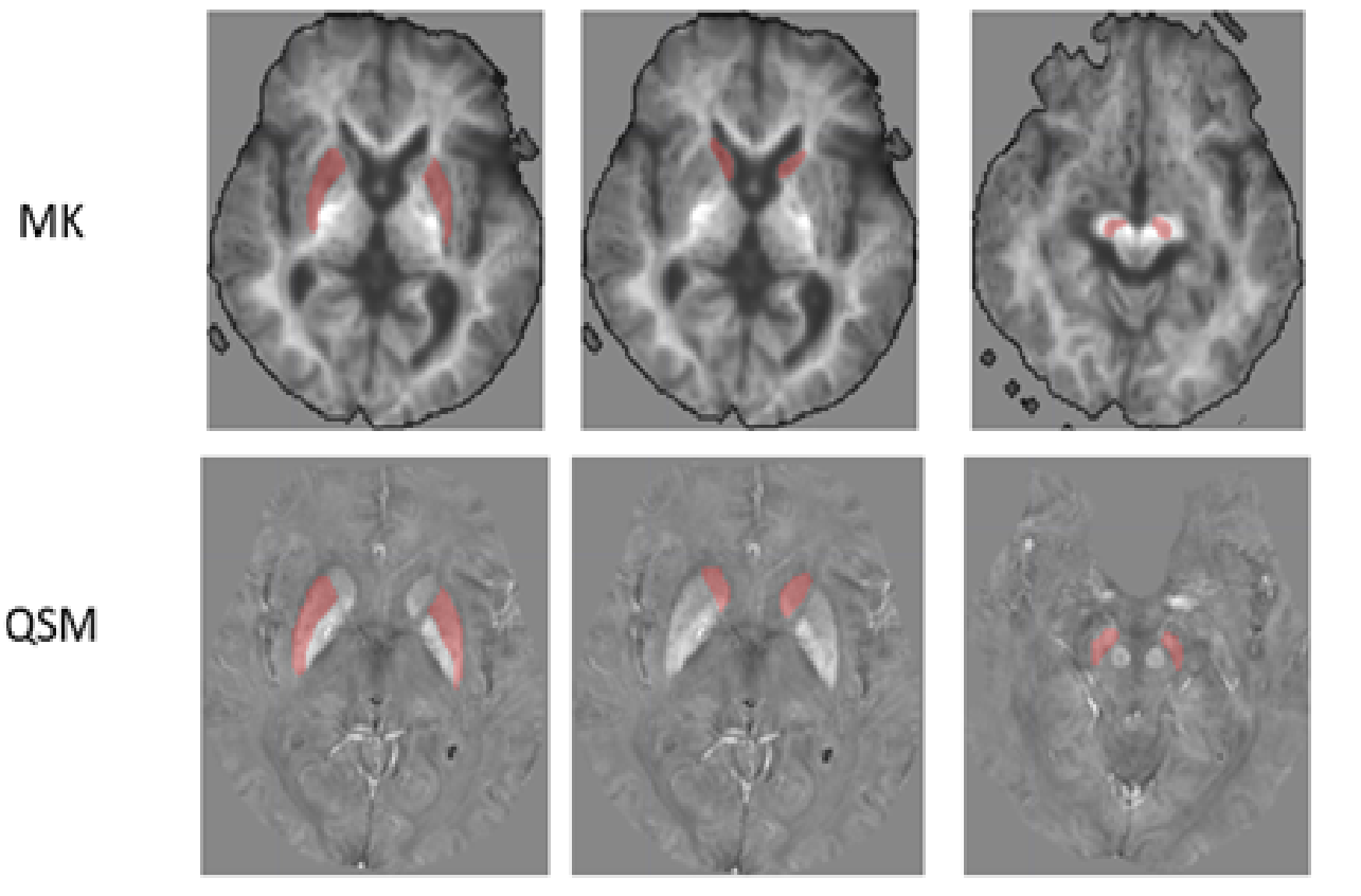
Method: Forty-seven PD patients (aged 68.7 years, 51% male, disease duration 0.78 years) and 16 healthy controls (aged 67.4 years, 63% male) were included. Participants underwent clinical evaluation using the Unified Parkinson’s Disease Rating Scale (MDS-UPDRS-3) and Hoehn & Yahr stage (H&Y), and 3T brain MRI including QSM and DKI. Regions-of-interest in the caudate nucleus, putamen, globus pallidus, and medial and lateral substantia nigra (SN) were manually drawn to compare the mean susceptibility (representing iron deposition) and DKI indices (representing restricted water diffusion) between PD patients and healthy controls (Figure 1).
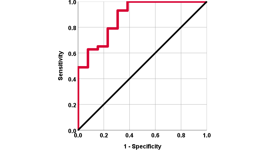
Results: Iron deposition was increased in PD in all ROIs except the caudate, and was significantly different after multiple comparison correction in the putamen (Figure 2; PD: 64.75 ppb, HC: 44.61 ppb, p=0.004). MD was significantly higher in PD in the lateral SN, putamen and caudate, the regions with the lowest iron deposition. In PD patients, we found significant association between the MDS-UPDRS-3 score and iron deposition in the putamen after correcting for age and sex (β=0.21, p=0.003). A composite DKI-QSM diagnostic marker based on these findings successfully differentiated the groups (p<0.0001) and had “good” classification performance (AUC=0.88; Figure 3).
Conclusion: QSM and DKI are complementary tools allowing better understanding of the complex interplay of iron deposition and microstructural changes in the pathophysiology of PD.
To cite this abstract in AMA style:
T. Welton, S. Hartono, S. Tan, YC. Shih, W. Lee, S. Chong, S. Ng, N. Chia, EK. Tan, L. Tan, LL. Chan. A combined imaging based biomarker for Parkinson’s disease using diffusion kurtosis and quantitative susceptibility mapping [abstract]. Mov Disord. 2021; 36 (suppl 1). https://www.mdsabstracts.org/abstract/a-combined-imaging-based-biomarker-for-parkinsons-disease-using-diffusion-kurtosis-and-quantitative-susceptibility-mapping/. Accessed April 19, 2025.« Back to MDS Virtual Congress 2021
MDS Abstracts - https://www.mdsabstracts.org/abstract/a-combined-imaging-based-biomarker-for-parkinsons-disease-using-diffusion-kurtosis-and-quantitative-susceptibility-mapping/


