Objective: To report a case of hemiballism associated with temporal lobe infarction.
Background: Hemiballism is one of the most striking hyperkinesias seen in clinical practice. It is usually associated with contralateral structural lesions in the subthalamic nucleus, other basal ganglia or even the contralateral cortex. Ipsilateral lesions have also been related to this abnormal movement and in some series about one third of cases have no abnormalities on neuroimaging.
Method: We report an 82-year-old right-handed woman with no previous medical history. A couple of weeks before admission to our service she was found on the floor of the kitchen by a family member. She spontaneously recovered in a few minutes and the event was considered as a syncope. During that day and the followings she complained of mild frontal headache. Because of persistent headache she visited a doctor who ordered some exams that weren’t performed. Three days before admission to our service she developed involuntary movements in the left side of the body. One of her sons noticed her somewhat disorientated with memory impairment and difficulties recognizing close relatives.
On admission, the neurological examination revealed moderate left hemiballism and mild hemiparesis in the left side with extensor plantar response. Consciousness was preserved. Bedside neuropsychological testing showed a mild amnestic syndrome with preserved executive functions.
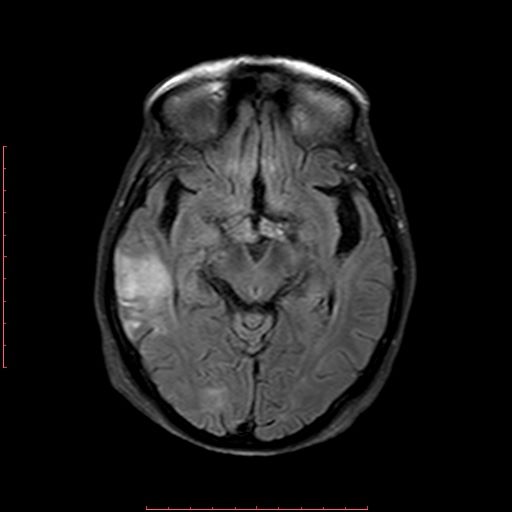
Results: Arterial hypertension and mild hypothyroidism were diagnosed and treated. Echocardiogram revealed mitral valvulopathy. No atrial fibrillation. CT scan showed a hypodensity of right temporal lobe. Brain MRI showed an acute ischemic stroke in the right temporal lobe [Figure 1]. An angiotomography revealed moderate stenosis of the right middle cerebral artery. Small dose of haloperidol was administered with adequate control of hemiballism in the subsequent days
Conclusion: Stroke, infections and metabolic disorders are the main etiologies of hemiballism. In a review of the literature we found a few older series reporting very scarce cases of ballism associated to, but not necessarily attributed to, a temporal and/or parietal ischemic lesions. It should be mentioned that CT scan was the most used neuroimaging study at that time.
To the best of our knowledge this could be the first case of the association of hemiballism and contralateral temporal lobe infarction as the only structural temporally–related demonstrable lesion.
To cite this abstract in AMA style:
B. Crispin, E. Mattos, R. Suarez, Y. Nuñez, C. Cosentino. A Case of Hemiballism Associated with Temporal Lobe Infarction [abstract]. Mov Disord. 2020; 35 (suppl 1). https://www.mdsabstracts.org/abstract/a-case-of-hemiballism-associated-with-temporal-lobe-infarction/. Accessed April 20, 2025.« Back to MDS Virtual Congress 2020
MDS Abstracts - https://www.mdsabstracts.org/abstract/a-case-of-hemiballism-associated-with-temporal-lobe-infarction/

