Category: Neuroimaging (Non-PD)
Objective: Evaluate cortical thickness in Huntington’s disease (HD) using 7 Tesla (T) MRI.
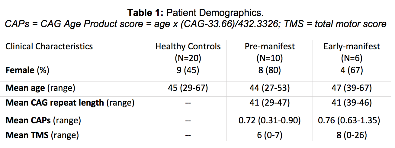
Background: There are currently no effective disease-modifying therapies for HD. In order to test the efficacy of putative treatment, robust and accurate methods that can reliably characterize disease extent and longitudinal progression prior to the onset of symptoms need to be validated. Cortical deterioration is thought to be an early pathological hallmark of HD progression together with striatal atrophy1, however, cortical changes have been poorly characterized to date. Compared to low-field MRI, 7T MRI can achieve superior anatomical resolution, resulting in more accurate delineation of brain structures and enhanced sensitivity to microstructural changes.2 In this study, therefore we exploited the benefits of 7T MRI to relate cortical thickness in 10 pre-manifest (PM) and 6 early-manifest (EM) HD patients, to clinical data and imaging data from 20 age-matched HC (Table 1).
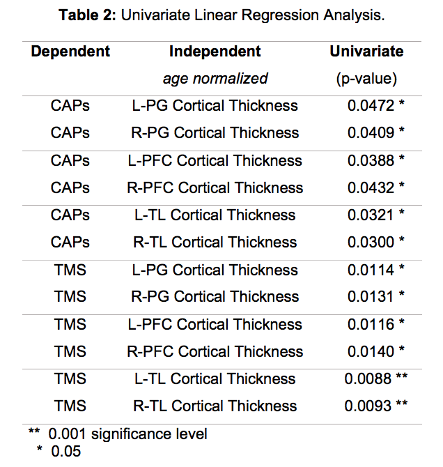
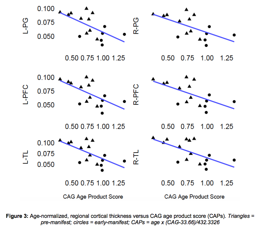
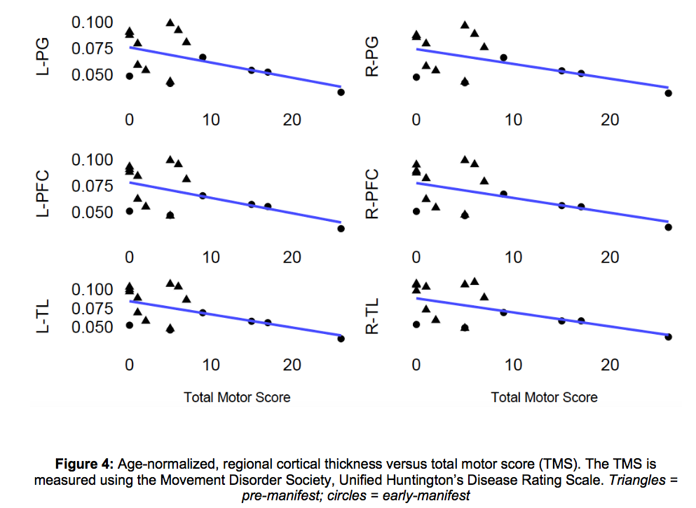
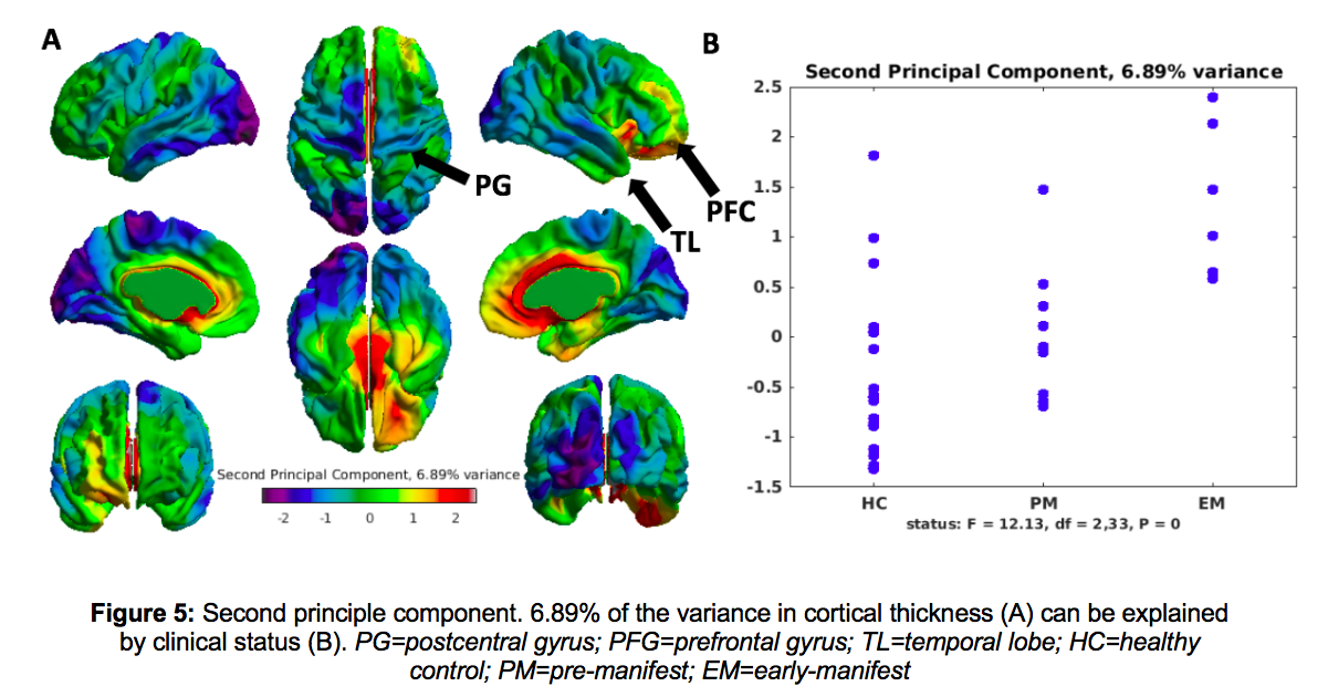
Method: Cortical thickness measurements were extracted from anatomical images using a robust brain morphology pipeline3,4. Regional thickness was quantified within the bilateral postcentral gyri (PG), prefrontal cortex (PFC), and temporal lobes (TL), based on a prior knowledge of their involvement in HD progression.5 Between-group comparisons were made. Regional thickness was associated with total motor scores (TMS) matched to the imaging timepoints, as well as the CAG-Age Product score (CAPs),6 a predictor of HD motor onset, using linear regression analysis. Independent component analysis (ICA) was used to explore sources of variance in whole-brain thickness across all groups.
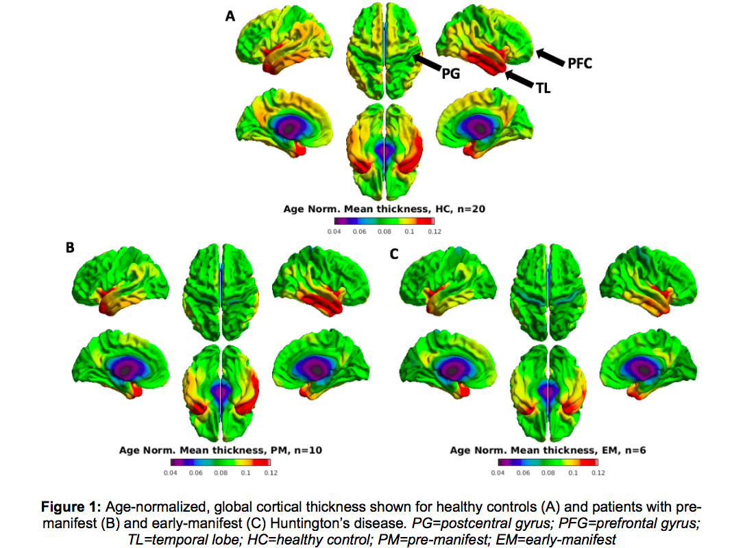
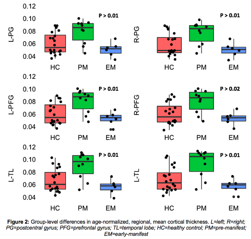
Results: Notable differences in age-normalized cortical thickness were observed between patients and controls, and amongst PM versus EM patients (Figs 1, 2). Regional thickness within the PG, PFC and TL, were significantly associated with CAPs and TMS, whereby, lower clinical scores were associated with thinning cortex (Table 2; Figs 3, 4). ICA revealed that after age (which accounts for 50.5% of the variance), 6.89% was explained by clinical status (HC, PM or EM) and 5.32% by sex differences.
Conclusion: Cortical thickness can help distinguish PM and EM patients with varying degrees of motor impairment, and is related to CAPs. Ongoing longitudinal studies in this cohort will help discern how early cortical changes can be identified in the PM gene-carriers.
References: 1. Sampedro F, Martinez-Horta S, Perez-Perez J, et al. Widespread Increased Diffusivity Reveals Early Cortical Degeneration in Huntington Disease. 2019. Am J Neuroradiol. 40(9):1464-1468. 2. Trattnig S, Springer E, Bogner W, et al. Key clinical benefits of neuroimage at 7T. 2016. NeuroImage. 168: 477-489. 3. Leynes AP, Morrison M, Jakary A, Larson P, Lupo J. 5221 Robust Bias Correction and Segmentation of 7 Tesla Structural Brain Images with an iterative Bias-Corrected Fuzzy C-means and N4 Bias Correction (iBCFCM+N4). Proc. Intl. Soc. Mag. Reson. Med. 25 2017. 4. Ad-Dab’bagh Y, Lyttelton O, Muehlboeck JS, et al. The CIVET image-processing environment: a fully automated comprehensive pipeline for anatomical neuroimaging research. 2006. Proceedings of the 12th Annual Meeting of the Organization of Human Brain Mapping. 5. Halliday GM, McRitchie DA, Macdonald V, et al. Regional specificity of brain atrophy in Huntington’s disease. 1998. Exp Neurol.154(2):663-672. 6. Hosung K, Ji-hoon K, Possin KL. Surface-based morphometry reveals caudate subnuclear structural damage in patients with premotor Huntington disease. 2017. Brain Imaging Behav. 11(5): 1365-1372.
To cite this abstract in AMA style:
M. Morrison, A. Jakary, S. Avadiappan, J. Tallakson, J. Glueck, A. Nelson, K. Possin, C. Dietiker, M. Geschwind, C. Hess, L. Lupo. 7 Tesla MRI investigation of cortical thickness in patients with Huntington’s disease and age-matched healthy controls [abstract]. Mov Disord. 2020; 35 (suppl 1). https://www.mdsabstracts.org/abstract/7-tesla-mri-investigation-of-cortical-thickness-in-patients-with-huntingtons-disease-and-age-matched-healthy-controls/. Accessed December 23, 2025.« Back to MDS Virtual Congress 2020
MDS Abstracts - https://www.mdsabstracts.org/abstract/7-tesla-mri-investigation-of-cortical-thickness-in-patients-with-huntingtons-disease-and-age-matched-healthy-controls/

