Session Information
Date: Sunday, October 7, 2018
Session Title: Parkinsonism, MSA, PSP (Secondary and Parkinsonism-Plus)
Session Time: 1:45pm-3:15pm
Location: Hall 3FG
Objective: We investigated the brain dopaminergic system and glucose metabolism in multiple system atrophy (MSA) patients with cognitive impairment.
Background: Since the first report of dementia in MSA, cognitive impairment in MSA patients has been increasingly recognized. Contrary to dementia in PD or DLB, cognitive deficits in MSA patients are characterized by short term memory loss and paucity of visual hallucinations. Recent pathological studies reported frequent globular NCIs (G-NCIs) in the medial temporal region, especially in their subiculum in patients with MSA and dementia.
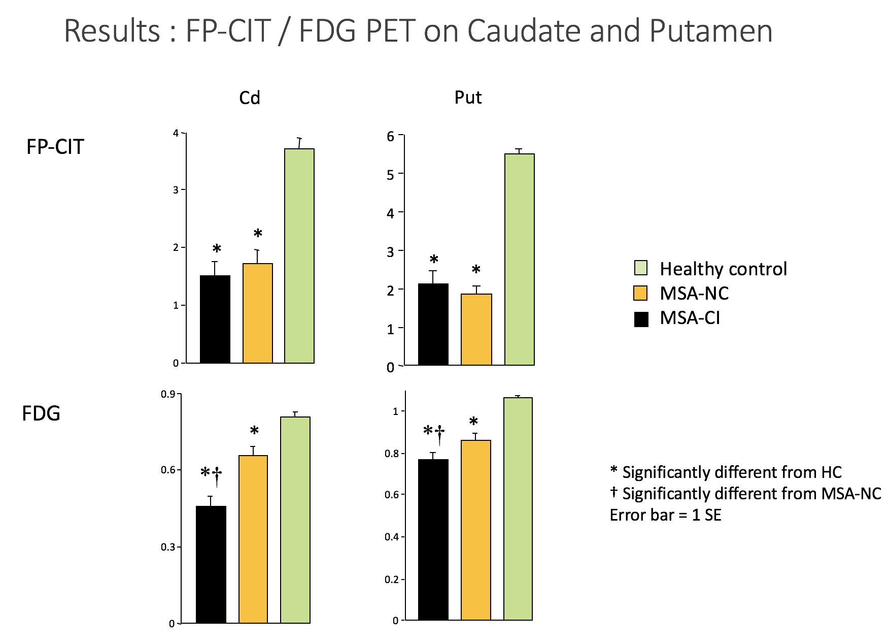
Methods: We studied 10 MSA patients with cognitive impairment (MSA-CI), 12 MSA with normal cognition (MSA-NC), and 19 age-matched healthy control subjects using [18F]FDG PET and [18F]FP-CIT PET. Severity of striatonigral degeneration in the two MSA groups were matched with FP-CIT binding in the putamen. Cognitive impairment in the MSA-CI group was assessed using the Mini-Mental State Examination and detailed neuropsychologic assessment. Glucose metabolism is expressed as metabolic ratio using V1 region as reference. FP-CIT binding is expressed as specific binding ratio using occipital cortex as reference.
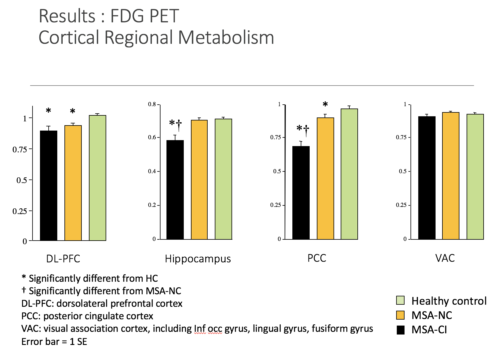
Results: Compared to the MSA-NC and healthy control groups, the MSA-CI group showed significant hypometabolism in the following regions: the insular, olfactory gyrus, posterior cingulate cortex, hippocampus, and the caudate nucleus. There was no significant difference in metabolism of visual association cortex. FP-CIT PET showed similar findings in the MSA-CI group: significant reduction of FP-CIT binding in the caudate and putamen. Exceptionally, 1 MSA cerebellar type patient with dementia showed normal FP-CIT PET findings.
Conclusions: We showed significant hypometabolism in the hippocampus, insula and caudate nucleus in MSA patients with cognitive impairment. These findings, consistent with recent pathological observations, suggest that dysfunction of the caudate nucleus and medial temporal regions may be related to the cognitive impairment of patients with MSA. Furthermore, our observations that cognitive impairment appears even in pure MSA-C patients, and the absence of correlation between striatal DA terminal loss and hypometabolism in the caudate and hippocampus indicate that cognitive impairment in MSA is independent of DA cell loss.
References: Kim HJ, Jeon BS, Kim YE, et al. Clinical and imaging characteristics of dementia in multiple system atrophy. Parkinsonism & related disorders. 2013;19(6):617–621.
To cite this abstract in AMA style:
KB. Lee, YJ. Park, C. Lee. [18F]FP-CIT and [18F]FDG PET findings in MSA patients with cognitive impairment [abstract]. Mov Disord. 2018; 33 (suppl 2). https://www.mdsabstracts.org/abstract/18ffp-cit-and-18ffdg-pet-findings-in-msa-patients-with-cognitive-impairment/. Accessed April 18, 2025.« Back to 2018 International Congress
MDS Abstracts - https://www.mdsabstracts.org/abstract/18ffp-cit-and-18ffdg-pet-findings-in-msa-patients-with-cognitive-impairment/


