Category: Choreas (Non-Huntington's Disease)
Objective: In Neuroacanthicytosis,to the best of our knowledge, bilateral fronto-temopral lobar atrophy associated with neuroacanthocytosis has not been reported. We report herein a case of chorea-acanthocytosis showing bilateral fronto-temporal atrophy with atrophy and signal intensity changes of striata on MR imaging.
Background: Neuroacanthocytosis is group of genetically heterogeneous disorders characterized by association of involuntary choreiform movements with peripheral blood acanthocytosis. Neuroimaging of neuroacanthocytosis shows atrophy of the striata, particularly the caudate nucleus, and increased signal intensity from the striata on T2-weighted imaging.
Method: we report a case of Neuroacanthocytosis.
Results: A 29-year-old man presented with involuntary movements of tongue and face for last 1.5 year. Chorea of the trunk and extremities had been present for past 1 year. He had history of seizure for last 10 years. Over past 2 years he had gradual decline of cognition involving memory and language domains. He had difficulty in speaking for last 1 year and he is not able to speak for last 6 months. The parents had non consanguineous marriage and one of his brother had involuntary face and body movements with seizures.
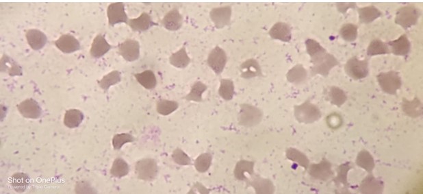
On general examination orofaciolingual dyskinesia and choreform involuntary movements of the face, trunk, and occasionally the extremities was present. There was mild generalized muscle atrophy with 4+ power in all limbs with hypotonia and hyporeflexia. Patient required support of two people to walk. On laboratory examination peripheral blood smear revealed abundant acanthocytes (Figure 2). Nerve conduction study showed evidence of bilateral sensorimotor axonopathy.
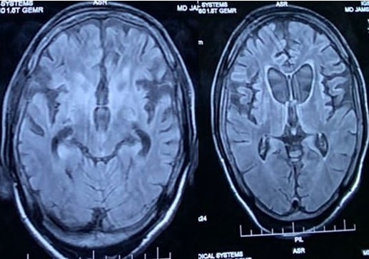
MR imaging examination of the brain revealed atrophy of the bilateral caudate nuclei and putamen with signal hyperintensity of the bilateral atrophic caudate and putamina on T2-weighted imaging. There was also marked fronto-temporal lobe atrophy. (Figure 1).
Conclusion: T In addition to atrophy and striatal signal intensity changes as a characteristic finding of neuroacanthocytosis, fronto-temporal atrophy may be seen as a spectrum of imaging findings in Neuroacanthocytosis.
Peripheral blood smear showing acanthocytes
FLAIR MR images showing fronto-temporal atrophy
To cite this abstract in AMA style:
N. Ranjan, AM. Kumar, AB. Ranjan. Bilateral Fronto-Temporal Lobar Atrophy: An Atypical Magnetic Resonance Imaging Finding in Neuroacanthocytosis [abstract]. Mov Disord. 2024; 39 (suppl 1). https://www.mdsabstracts.org/abstract/bilateral-fronto-temporal-lobar-atrophy-an-atypical-magnetic-resonance-imaging-finding-in-neuroacanthocytosis/. Accessed April 1, 2025.« Back to 2024 International Congress
MDS Abstracts - https://www.mdsabstracts.org/abstract/bilateral-fronto-temporal-lobar-atrophy-an-atypical-magnetic-resonance-imaging-finding-in-neuroacanthocytosis/


