Category: Surgical Therapy: Parkinson's Disease
Objective: The study evaluates the prediction capacity of two different Deep brain stimulation (DBS) electrode reconstructions methods: Lead-DBS and StimView . The methods predictions of the most potentially effective contacts were, then, compared based on clinical examination (standard of care).
Background: DBS programming is getting more challenging(1). Therefore, tools capable of optimize programming duration time are necessary(2,3,4).
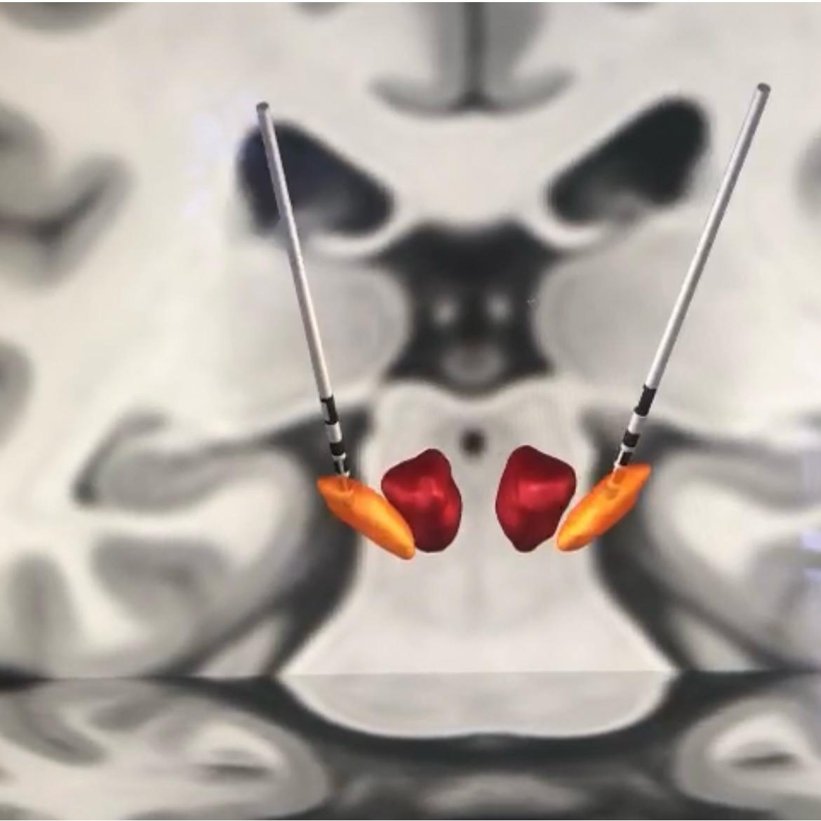
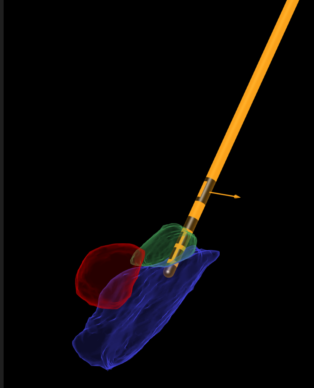
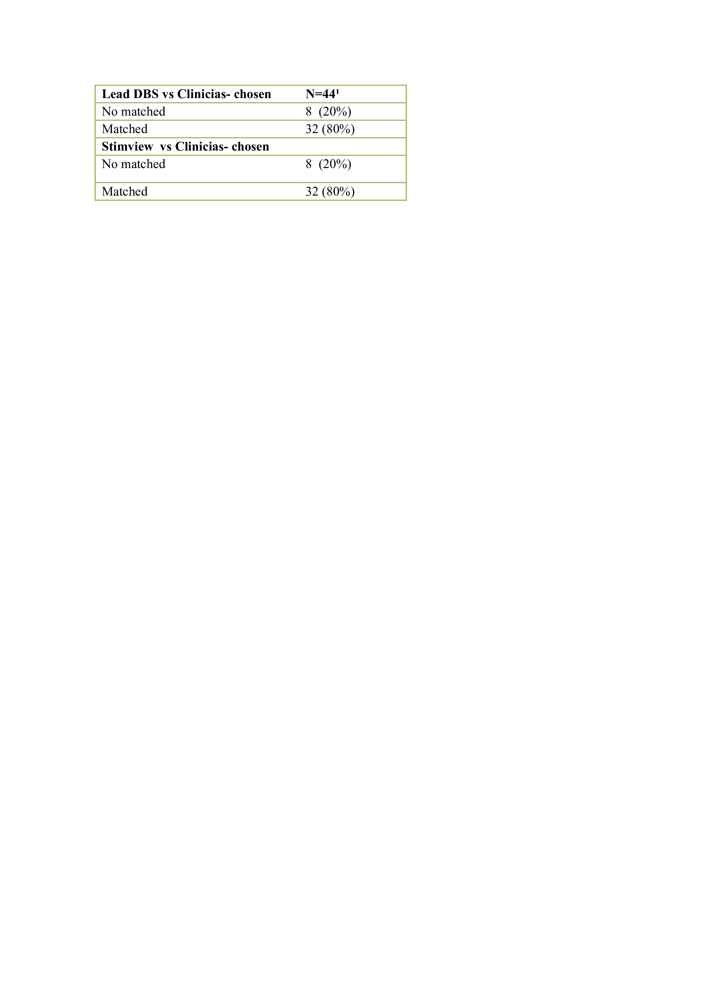
Method: 29 patients were included, 27 with Parkinson`s disease (PD) and 2 with dystonia (DT). Therefore, 58 brain hemispheres images were then obtained. Of these, 56 had directional leads and 2 had leads with eight ring contacts. Lead-DBS and StimView use preoperative magnetic resonance image (MRI) fused with postoperative computed tomography (CT). (2,5) The same images series were imported into the softwares, then fused and their accuracy verified (2,5). Some differences exist between those two methods as Lead-DBS tool works with a normalized image (2), which does not occur in StimView process (5). The anatomic relationship between the electrodes and the subthalamic nucleus (STN) and internal globus pallidus (GPi) were analyzed (Figure 1). The best contacts are usually in the vicinity of known regions for motor symptoms improvement. For PD, this area corresponds to the dorsolateral portion the STN (6) and posterodorsal portion of the GPi (7). In DT cases, is located in the posterior ventro-lateral portion of the GPi (8). Based on the reconstructed image, the closest contact level to these areas was chosen. Also, if the contacts levels were directional contacts, the best located directional contacts were also chosen (44/58 it was possible). The best contact was defined as the one with the highest motor symptoms improvement rate based on clinical exam. Inferential statistics were performed using the chi-square association, having a significance level of p<0.05.
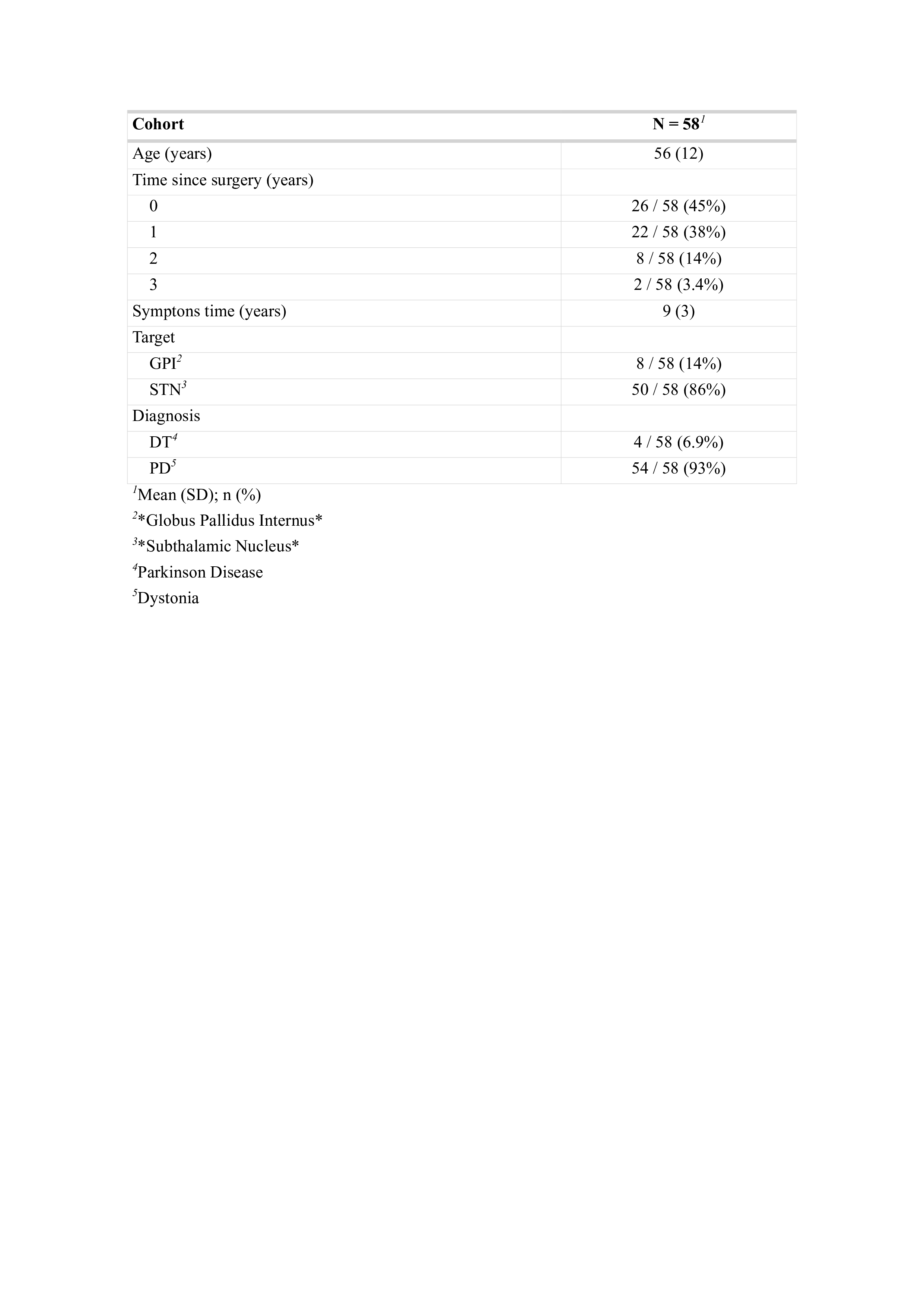
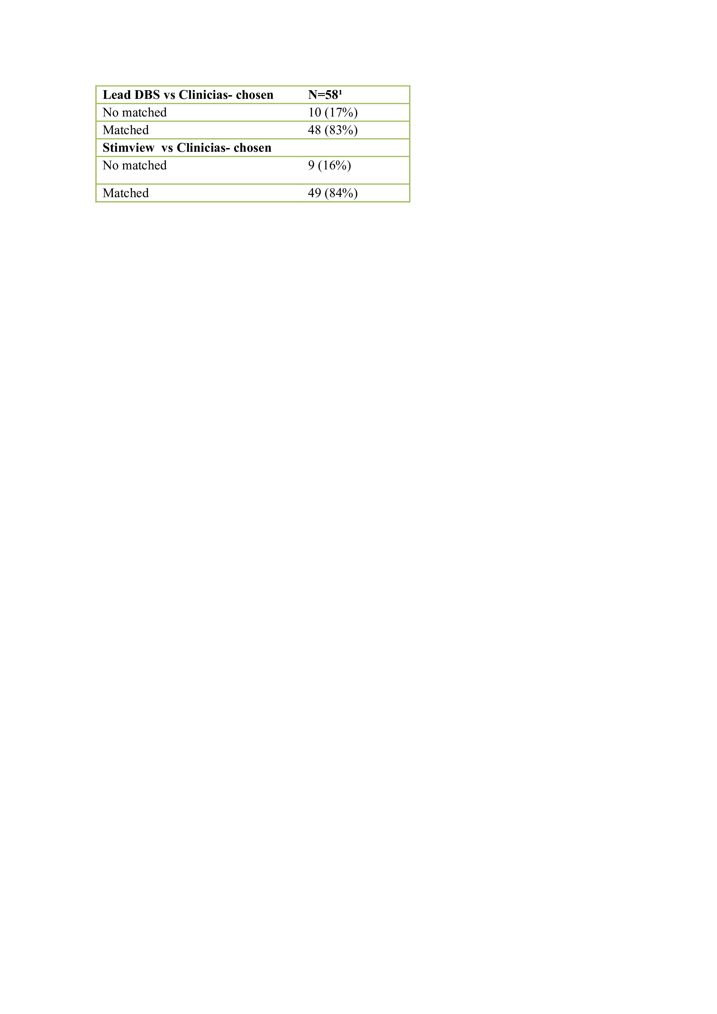
Results: The mean age was 56 and 24 patients had surgery in a year or less -the maximum time was three years as table 1. The Lead-DBS was capable to predict 48/58 of the clinician-chosen contacts and the Stim-View predicted 49/58 as table 2 (x² 8.012 and p-value= 0.004647). The prediction capacity of directional contacts were 80 % in both methods as table 3 (x² 14.854 and p-value=0.0001162).
Conclusion: Both methods seemed effective in image prediction.
Figure 1A:The reconstructed Lead DBS
Figure 1B:The reconstructed StimView
Table1: Participant characteristics
Table 2
Table 3
References: 1. Xu SS, Lee WL, Perera T, et al. Can brain signals and anatomy refine contact choice for deep brain stimulation in Parkinson’s disease? [published online ahead of print, 2022 May 19]. J Neurol Neurosurg Psychiatry. 2022;jnnp-2021-327708.
2. Horn A, Li N, Dembek TA, et al. Lead-DBS v2: Towards a comprehensive pipeline for deep brain stimulation imaging. Neuroimage. 2019;184:293-316.
3. Jaradat A., Nowacki A., Montalbetti M., Debove I., Petermann K., Schlaeppi J.-A., Lachenmayer L., Tinkhauser G., Krack P., Nguyen T.-A.K., Pollo C. 2023. Probabilistic Subthalamic Nucleus Stimulation Sweet Spot Integration Into a Commercial Deep Brain Stimulation Programming Software Can Predict Effective Stimulation Parameters. Neuromodulation 2023; 26: 348–355.
4. Xu, Y., Qin, G., Tan, B., Fan, S., An, Q., Gao, Y., Fan, H., Xie, H., Wu, D., Liu, H., Yang, G., Fang, H., Xiao, Z., Zhang, J., Zhang, H., Shi, L., & Yang, A. (2023). Deep Brain Stimulation Electrode Reconstruction: Comparison between Lead-DBS and Surgical Planning System. Journal of clinical medicine, 12(5), 1781.
5. Lange F, Steigerwald F, Malzacher T, et al. Reduced Programming Time and Strong Symptom Control Even in Chronic Course Through Imaging-Based DBS Programming. Front Neurol. 2021;12:785529. Published 2021 Nov 8.
6. Dembek TA, Roediger J, Horn A, et al. Probabilistic sweet spots predict motor outcome for deep brain stimulation in Parkinson disease. Ann Neurol. 2019;86(4):527-538.
7. Wong JK, Hilliard JD, Holanda VM, et al. Time for a New 3-D Image for Globus Pallidus Internus Deep Brain Stimulation Targeting and Programming. J ParkinsonsDis. 2021;11(4):1881-1885. doi:10.3233/JPD-212820
8. Reich MM, Horn A, Lange F, et al. Probabilistic mapping of the antidystonic effect of pallidal neurostimulation: a multicentre imaging study. Brain. 2019;142(5):1386-1398.
To cite this abstract in AMA style:
R. Barbosa, J. Yamamoto, M. Soares, I. Paraguay, S. Casagrande, J. Dornelly, M. Bezerra, G. Torezani, V. Milanese, G. Micheli, E. Barbosa, R. Cury. Comparison of two methods of deep brain stimulation lead reconstruction and their prediction capacities [abstract]. Mov Disord. 2024; 39 (suppl 1). https://www.mdsabstracts.org/abstract/comparison-of-two-methods-of-deep-brain-stimulation-lead-reconstruction-and-their-prediction-capacities/. Accessed October 21, 2025.« Back to 2024 International Congress
MDS Abstracts - https://www.mdsabstracts.org/abstract/comparison-of-two-methods-of-deep-brain-stimulation-lead-reconstruction-and-their-prediction-capacities/

