Category: Parkinson's Disease: Neuroimaging
Objective: To elucidate regional neural activity differences in patients with PD-FOG using resting-state fMRI (rs-fMRI).
Background: Freezing of gait (FOG) in Parkinson’s disease (PD0 is an issue with gait where those affected are unable to begin walking, turn, or move around in tight spaces. Little is known about why freezing of gait occurs, although one theory behind why FOG happens is an alteration in neural connectivity between certain areas of the brain. Neuronal activity causes low-frequency fluctuations of brain activity which can be measured by (ALFF) and its standardized counterpart, fractional ALFF (fALFF). We hypothesize that we will see decreased ALFF values and increased fALFF values in those with PD-FOG compared to PD-nFOG and HC.
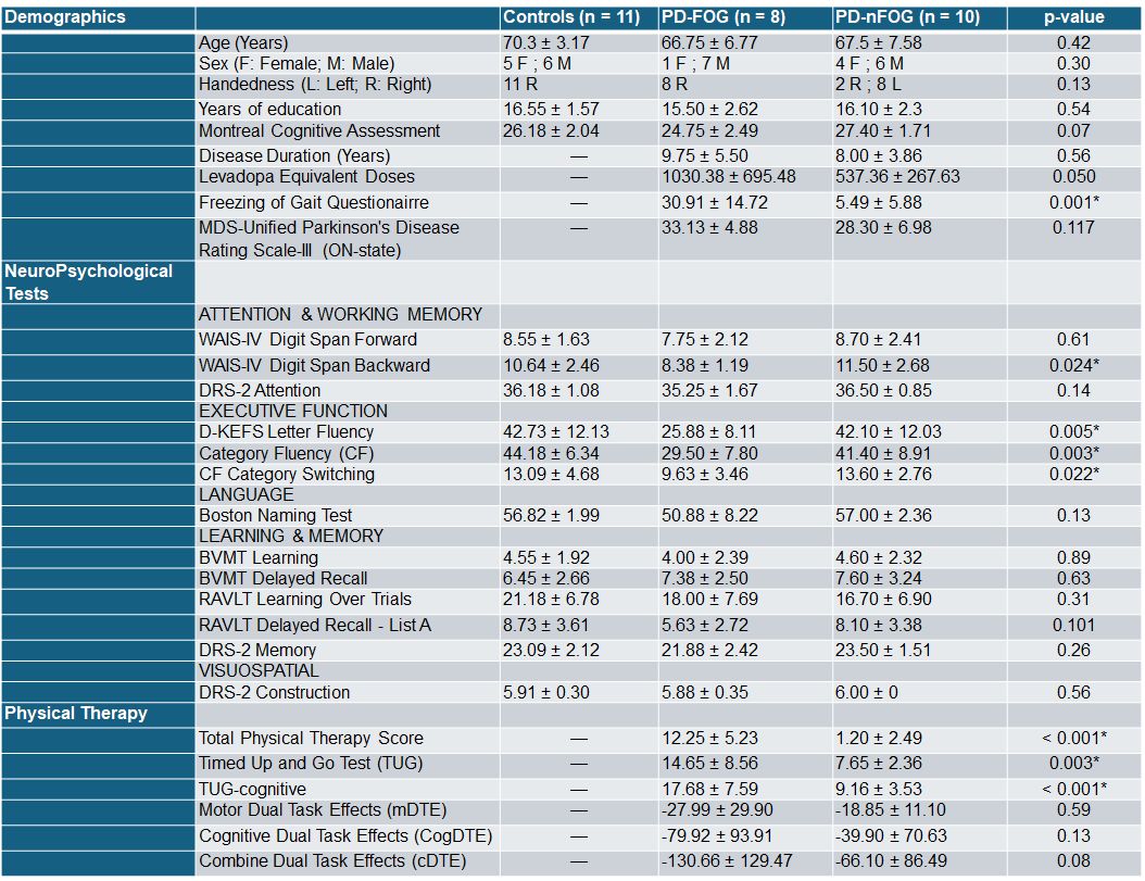
Method: 8 patients with PD-FOG, 10 with PD-nFOG, and 11 healthy controls (HC) were recruited for this study. All subjects underwent rs-fMRI scans on a 3T Siemens Skyra scanner. The rs-fMRI involved gradient-echo T2*-weighted echo-planar imaging (EPI) acquisition (repetition time [TR] = 700 ms, echo time [TE] = 28.4 ms, flip angle = 42°, resolution = 2.3 × 2.3 × 2.3 mm3, 64 axial slices, multiband = 8). All BOLD data underwent preprocessing using fMRIB Software Library (FSL; https://fsl.fmrib.ox.ac.uk/fsl/fslwiki), Advanced Normalization Tools (ANTs; https://github.com/ANTsX/ANTs/wiki), and in-house MATLAB scripts. We obtained general demographics for all subjects and the disease duration, FOGQ score, levodopa equivalent doses (LEDD), physical therapy tests ([table 1]) , and MDS-UPDRS-III scores for each patient with PD. We collected neuropsychological tests scores for all participants ([table 1]).
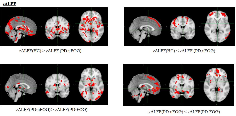
Results: We found trending differences (p<0.05) in ALFF and fALFF between HC and PD-nFOG, HC and PD-FOG, and PD-nFOG and PD-FOG. Specifically, we found changes in ALFF values between HC and PD-nFOG in the frontoparietal and occipital regions and changes in ALFF between PD-nFOG and PD-FOG in the frontoparietal regions. We also found changes in fALFF values between HC and PD-FOG in the frontoparietal regions (not shown).
Conclusion: Our analysis concluded that there are differences in regional activity of areas involved in visuospatial perception and movements, which can be measured by ALFF and fALFF. Future analysis of the correlation with neuropsychological scores is underway.
zALFF maps
Demographics
To cite this abstract in AMA style:
T. Davis, Z. Mari, V. Mishra. Differences in regional activity levels between healthy controls, PD-nFOG, and PD-FOG as measured by ALFF and fALFF using resting-state fMRI. [abstract]. Mov Disord. 2024; 39 (suppl 1). https://www.mdsabstracts.org/abstract/differences-in-regional-activity-levels-between-healthy-controls-pd-nfog-and-pd-fog-as-measured-by-alff-and-falff-using-resting-state-fmri/. Accessed April 1, 2025.« Back to 2024 International Congress
MDS Abstracts - https://www.mdsabstracts.org/abstract/differences-in-regional-activity-levels-between-healthy-controls-pd-nfog-and-pd-fog-as-measured-by-alff-and-falff-using-resting-state-fmri/


