Category: Parkinsonism, Atypical: PSP, CBD
Objective: We aim to investigate the relationship between regional 18F-florzolotau uptake and clinical severity, structural volume changes, and plasma markers in tauopathy parkinsonism syndrome.
Background: 18F-florzolotau positron emission tomography (PET) assists in the in vivo diagnosis of tauopathy parkinsonism syndromes, specifically progressive supranuclear palsy (PSP). However, the associations of this novel tau PET imaging and other available noninvasive biomarkers, the plasma biomarkers and structural imaging, are not fully elucidated.
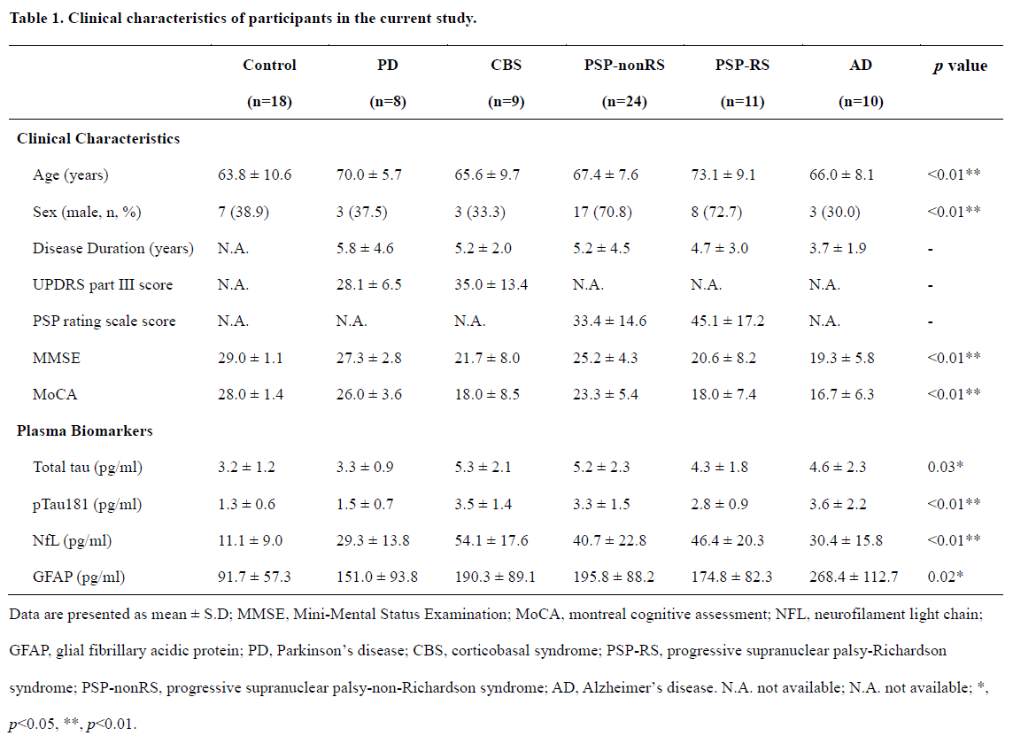
Method: A total of 80 participants were recruited: 11 with PSP-Richardson syndrome, 24 with PSP non-Richardson syndrome, 9 with corticobasal syndrome (CBS), 10 with Alzheimer’s disease (AD), 8 with Parkinson’s disease, and 18 age-matched healthy controls. All participants underwent 18F-florzolotau PET, brain MRI, and single-molecule array (Simoa)-based plasma biomarker investigation (total and phosphorylated tau [pTau181], neurofilament light chain, and glial fibrillary acidic protein [GFAP]).
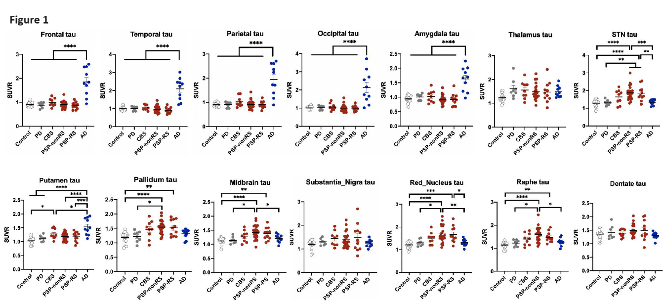
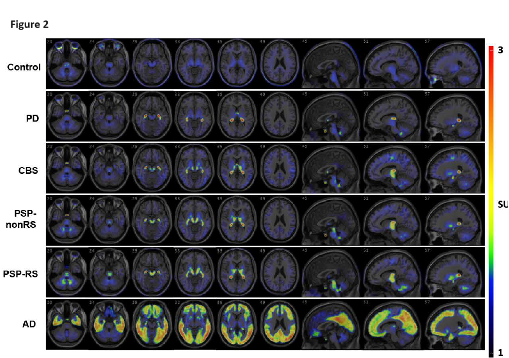
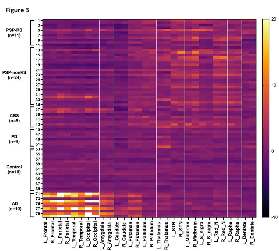
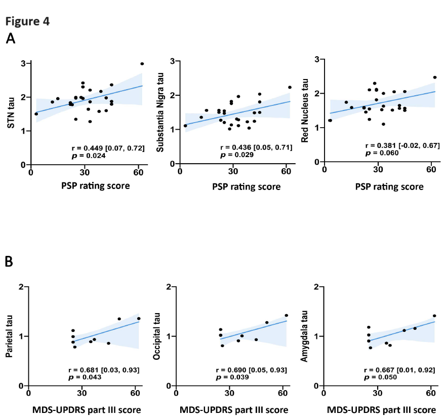
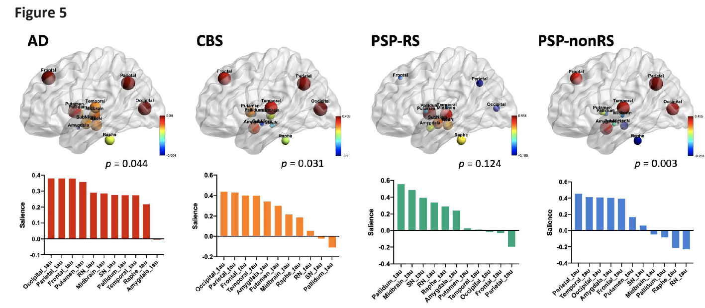
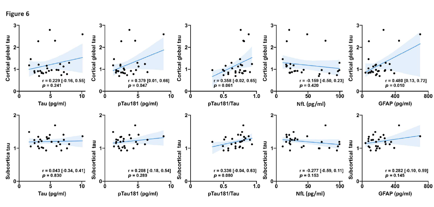
Results: 18F-florzolotau uptake was significantly higher in the subcortical regions of the pallidum, subthalamic nucleus, midbrain, red nucleus, and raphe nucleus in PSP patients compared to the other groups and controls (all p<0.01)(Figure 1-3). Subcortical tau tracer retention satisfactorily assisted in distinguishing PSP and CBS from controls (AUC=0.952, p<0.001) and AD (AUC=0.982, p<0.001). The severity of PSP positively correlated with tau burden in the above subcortical regions (Figure 4A). The motor severity of CBS correlated with tau retention in the parietal, occipital cortex, and amygdala (Figure 4B). Tau tracer uptake was associated with cortical volume changes in CBS (p=0.031), PSP non-Richardson syndrome (p=0.003), and AD (p=0.044)(Figure 5). Cortical tau retention correlated with plasma levels of GFAP (p=0.010) and pTau181 (p=0.047) in tauopathy disorders (Figure 6).
Conclusion: 18F-Florzolotau uptake in specific subcortical regions assists in the differential diagnosis of PSP and CBS. In addition, regional tau burden contributes to structural volume changes and correlates with plasma levels of GFAP and marginally pTau181.
Table 1
Figure 1
Figure 2
Figure 3
Figure 4
Figure 5
Figure 6
To cite this abstract in AMA style:
CH. Li, SP. Fan, MC. Shih, YH. Weng, TF. Chen, IT. Hsiao, KJ. Lin, CH. Lin. 18F-Florzolotau Positron Emission Tomography Imaging Correlates with Regional Brain Atrophy and Plasma Biomarkers in Tauopathy Parkinsonism Syndrome [abstract]. Mov Disord. 2024; 39 (suppl 1). https://www.mdsabstracts.org/abstract/18f-florzolotau-positron-emission-tomography-imaging-correlates-with-regional-brain-atrophy-and-plasma-biomarkers-in-tauopathy-parkinsonism-syndrome/. Accessed March 31, 2025.« Back to 2024 International Congress
MDS Abstracts - https://www.mdsabstracts.org/abstract/18f-florzolotau-positron-emission-tomography-imaging-correlates-with-regional-brain-atrophy-and-plasma-biomarkers-in-tauopathy-parkinsonism-syndrome/

