Category: Parkinsonism, Atypical: PSP, CBD
Objective: To investigate nigral neuroinflammatory markers and dopaminergic neuron counts in parkinsonian disorders, comparing their profiles across conditions.
Background: In degenerative parkinsonian disorders, the primary loss of dopaminergic neurons occurs in the substantia nigra pars compacta (SNc). Limited data exist on the involvement of neuroinflammation within the SNc, and comparative studies across the disorders are lacking. We investigated counts and densities of SNc dopaminergic neurons, neuroinflammatory T-cells, and microglial cells in PD, PSP and MSA, comparing them to healthy controls (HC) while exploring clinical characteristics.
Method: Postmortem neuropathological samples were obtained from 79 individuals (PD n=38, PSP n=15, MSA n=14, HC n=12). The numbers and densities of SNc tyrosine hydroxylase (TH)-positive neurons and T-cells (CD3+, CD4+ and CD8+), and Iba1 expression (indicative of microglia) in both the SNc and the crus cerebri were assessed. Demographic and clinical phenotypical data were collected from individual patient histories.
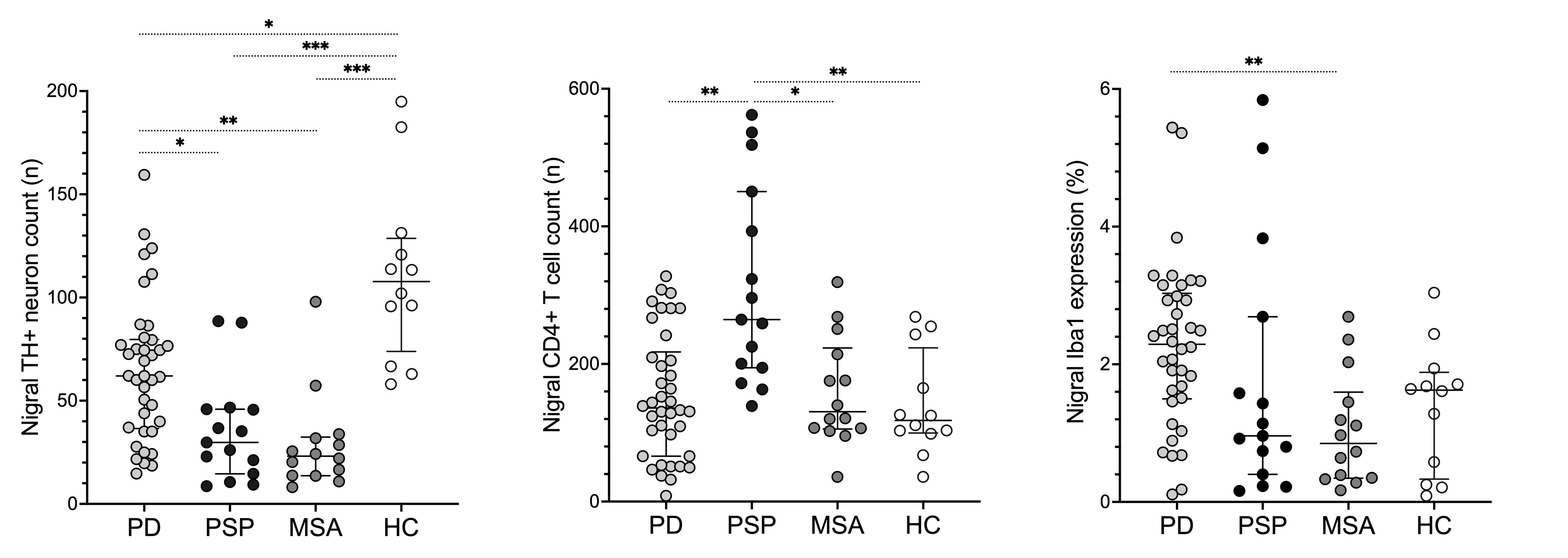
Results: PSP patients had substantially 89-212% higher counts of nigral CD3+, CD4+, and CD8+ T-cells compared to MSA patients (p<0.04), 125-178% higher counts of CD3+ and CD4+ T-cells compared to healthy controls (p<0.002), and a 95% increase in the counts of CD4+ T-cells compared to PD patients (p=0.001) [Figure 1]. Iba1 expression in the SNc was significantly elevated in PD patients compared to MSA patients (p=0.004), with no significant variations observed across other conditions. The counts of SNc TH+ neurons in PSP and MSA patients were 58-67% lower compared to PD patients (p<0.05) and 73-78% lower compared healthy controls (p<0.001). Nigral dopaminergic neuronal loss did not correlate with neuroinflammatory activity in any of the conditions.
Conclusion: PSP is associated with distictive T-cell-based nigral neuroinflammatory activity, while PD predominantly displays microglia-based neuroinflammation. Both MSA and PSP demonstrate severe losses of nigral dopaminergic neurons compared to PD. Notably, the neuroinflammatory response appears to be independent of the extent of dopaminergic neuronal loss in all conditions, indicating parallel but independent processes. Alternatively, the substantial increase of T-cells contributes to neuron loss, but the process involves currently unknown modifying factors.
Figure 1
To cite this abstract in AMA style:
E. Backman, M. Gardberg, L. Luntamo, M. Peurla, T. Vahlberg, P. Borghammer, N. Stefanova, G. Wenning, V. Kaasinen. Nigral Neuroinflammation and Dopaminergic Neurons in PD, MSA and PSP: A Comparative Clinicopathological Study [abstract]. Mov Disord. 2024; 39 (suppl 1). https://www.mdsabstracts.org/abstract/nigral-neuroinflammation-and-dopaminergic-neurons-in-pd-msa-and-psp-a-comparative-clinicopathological-study/. Accessed January 1, 2026.« Back to 2024 International Congress
MDS Abstracts - https://www.mdsabstracts.org/abstract/nigral-neuroinflammation-and-dopaminergic-neurons-in-pd-msa-and-psp-a-comparative-clinicopathological-study/

