Category: Dystonia: Pathophysiology, Imaging
Objective: We employed a multimodal approach to gain a comprehensive understanding of the pathophysiology of cervical dystonia. The aim was to assess behavioural, microstructural, and functional connectivity differences between patients, their first-degree relatives, and healthy controls.
Background: Dystonia is a complex and heterogeneous movement disorder. This has led to a gap in diagnoses, with limited objective measures available. The disease appears to affect the brain at a network level, not only diminishing motor function but also other neuroprocessing pathways [1]. Existing studies have already demonstrated behavioural processing level differences in dystonia patients [1], and recent machine learning approaches have demonstrated measurable microstructural differences [2]. The aim of this study was to combine these modalities with the addition of functional and resting state MRI data.
Method: This study collected data from 64 subjects, including cervical dystonia patients (16), first-degree relatives (16 abnormal, 16 normal), and age and gender-matched healthy controls (16). The data includes behavioural measures of Temporal Discrimination Thresholds (TDT), structural MRI, resting-state fMRI, and temporal discrimination-focused fMRI. A multivariate approach was used to incorporate information across structural, functional, and behavioural differences between the groups.
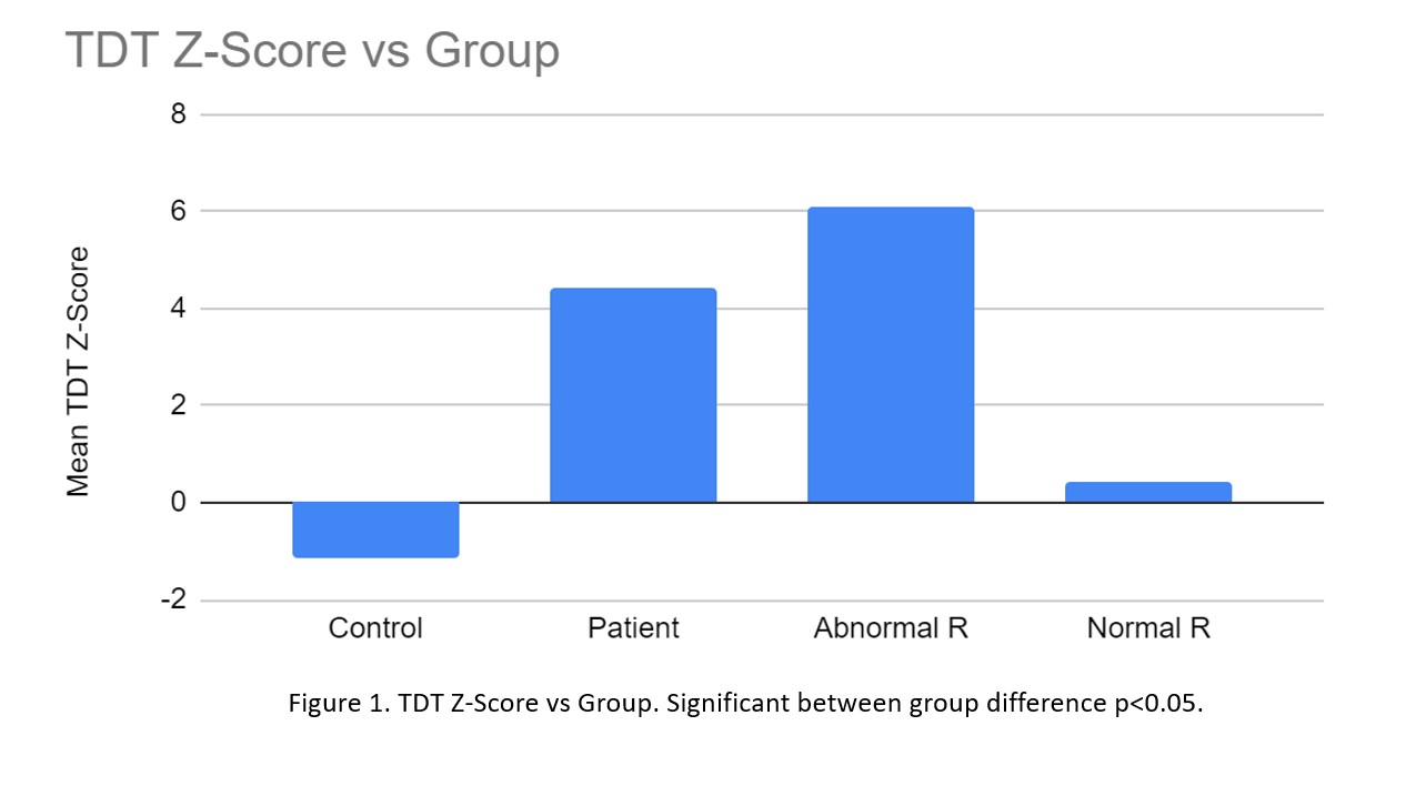
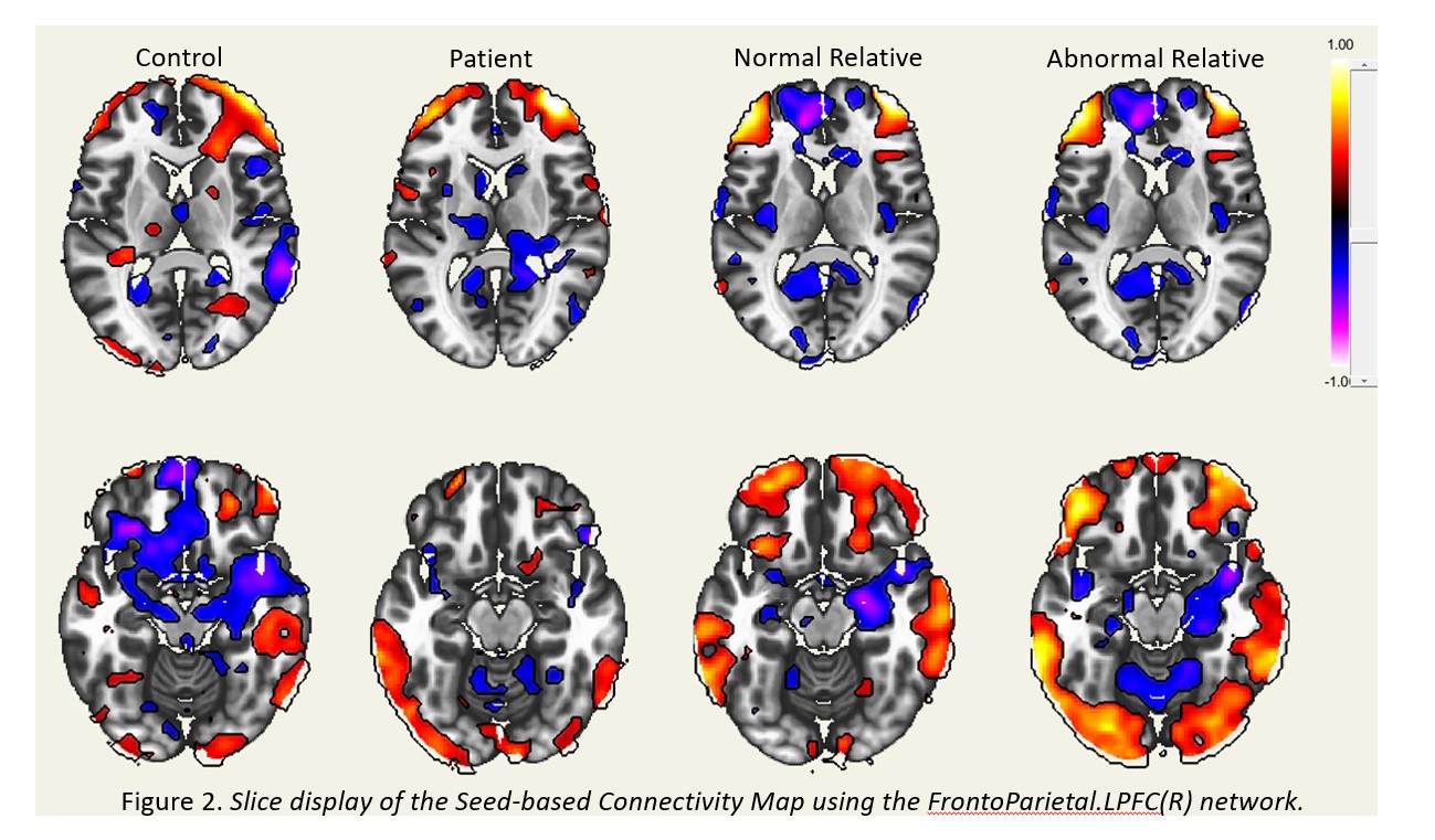
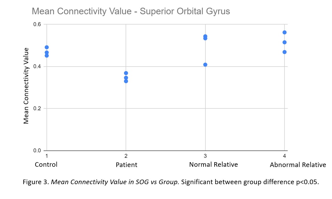
Results: Replicating previous results, significant differences in TDT values were demonstrated in an initial subset of our data [Figure 1]. The FrontoParietal network was used in seed-based connectivity analysis for our functional data [Figure 2]. This showed significant between group differences [Figure 3] in mean connectivity values. This network was used as it contains the superior orbital gyrus, previously connected to dystonia [2,3]. Connectivity in this region was not significantly correlated with TDT values (r 0.3, p>0.05). We plan to reveal further network-level variability between patients, relatives, and healthy controls which may account for behavioural differences seen at each group level.
Conclusion: This study demonstrates the validity of using a multimodal approach for modeling dystonia. The integration of structural, functional, and resting-state data provides a more holistic overview. The results suggest that microstructural differences may correlate with network-level differences reflected in functional data.
References: 1. Kimmich O, Molloy A, Whelan R, Williams L, Bradley D, Balsters J, Molloy F, Lynch T, Healy DG, Walsh C, O’Riordan S, Reilly RB, Hutchinson M. Temporal discrimination, a cervical dystonia endophenotype: penetrance and functional correlates. Mov Disord. 2014 May;29(6):804-11. doi: 10.1002/mds.25822. Epub 2014 Jan 30. PMID: 24482092.
2. Valeriani D, Simonyan K. A microstructural neural network biomarker for dystonia diagnosis identified by a DystoniaNet deep learning platform. Proc Natl Acad Sci U S A. 2020 Oct 20;117(42):26398-26405. doi: 10.1073/pnas.2009165117. Epub 2020 Oct 1. PMID: 33004625; PMCID: PMC7586425.
3. Wei S, Chen X, Xiao Y, Jiang W, Yin Q, Lu C, Yang L, Wei J, Liu Y, Li W, Tang J, Guo W, Luo S. Abnormal Network Homogeneity in the Right Superior Medial Frontal Gyrus in Cervical Dystonia. Front Neurol. 2021 Nov 5;12:729068. doi: 10.3389/fneur.2021.729068. PMID: 34803879; PMCID: PMC8602349.
To cite this abstract in AMA style:
SR. Bostan, R. King, C. Fearon, M. Hutchinson, R. Reilly. Multimodal neuroimaging to probe brain networks in Cervical Dystonia. [abstract]. Mov Disord. 2023; 38 (suppl 1). https://www.mdsabstracts.org/abstract/multimodal-neuroimaging-to-probe-brain-networks-in-cervical-dystonia/. Accessed February 2, 2026.« Back to 2023 International Congress
MDS Abstracts - https://www.mdsabstracts.org/abstract/multimodal-neuroimaging-to-probe-brain-networks-in-cervical-dystonia/


