Category: Parkinson's Disease: Neuroimaging
Objective: To detect the signal change of nigrosome-1 in 7T-MRI and its diagnostic and differential diagnostic efficacy as the imaging biomarker of PD, MSA-P, MSA-C and PSP.
Background: Current findings on the signal intensity change of nigrosome-1 in Parkinson’s disease patients and Parkinson-plus syndrome patients are inconsistent, due to limited number of participants in 7T-MRI study and different evaluation criteria for abnormal nigrosome-1 signal change. Thus, the characteristics and differences of nigrosome-1 lesions in patients with Parkinson’s disease (PD), multiple system atrophy (MSA) and progressive supranuclear palsy (PSP) still need further study.
Method: Healthy individuals and patients with PD, MSA, and PSP were recruited. All participants underwent 7T-MRI scans with 3D T2*-weighted imaging with multi-echo acquisition. Two physicians were invited to score the visibility of bilateral nigrosome-1 blindly and independently. The receiver operating characteristic (ROC) curve was used to evaluate the diagnostic accuracy.
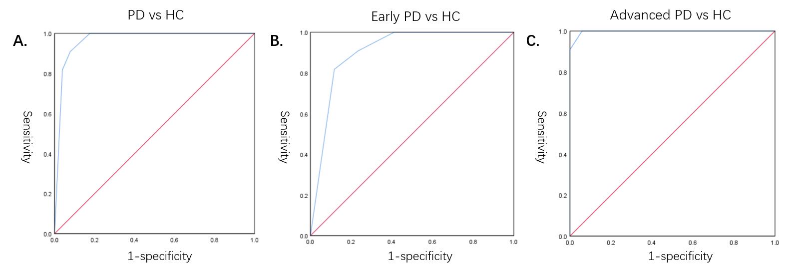
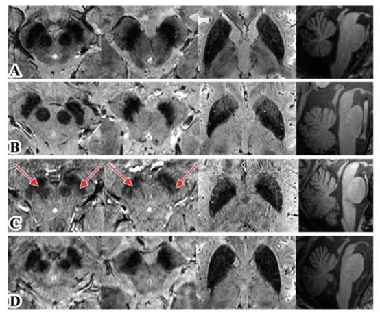
Results: A total of 51 PD patients, 45 MSA patients, 10 PSP patients, and 22 age- and sex-matched healthy controls were included. The nigrosome-1 score in the PD group was significantly lower than control group (P<0.001). The cut-off value of the nigrosome-1 score for distinguishing PD patients from HCs was 2.5, with sensitivity, specificity of 92.2%, 90.9%, respectively, the AUC was 0.967. The cut-off value of nigrosome-1 score in patients with early PD was 3.5, with sensitivity, specificity of 88.2%, 81.8%, respectively, the AUC was 0.906. The cut-off value of nigrosome-1 score in patients with advanced PD was 1.5, with sensitivity, specificity of 94.1%, 100%, respectively, the AUC was 0.997 (Figure 1). Interestingly, the nigrosome-1 score in the MSA-P and PSP group were significantly lower than the MSA-C group (P=0.006, P=0.048), no significant difference was found between other disease groups (Figure 2).
Conclusion: Nigrosome-1 signal change showed high sensitivity and specificity in distinguishing PD, MSA-P, MSA-C, and PSP patients from healthy individuals. The nigrosome-1 score in MSA-C patients was significantly higher than that in MSA-P and PSP patients, suggesting a relatively preserved nigrosome-1 hyperintensity in MSA-C patients.
References: 1 BLAZEJEWSKA A I, SCHWARZ S T, PITIOT A, et al. Visualization of nigrosome 1 and its loss in PD: pathoanatomical correlation and in vivo 7 T MRI [J]. Neurology, 2013, 81(6): 534-40.
2 POSTUMA R B, BERG D, STERN M, et al. MDS clinical diagnostic criteria for Parkinson’s disease [J]. Movement disorders : official journal of the Movement Disorder Society, 2015, 30(12): 1591-601.
3 LANCIONE M, DONATELLI G, DEL PRETE E, et al. Evaluation of iron overload in nigrosome 1 via quantitative susceptibility mapping as a progression biomarker in prodromal stages of synucleinopathies [J]. NeuroImage, 2022, 260: 119454.
To cite this abstract in AMA style:
D. Su, Z. Zhang, Z. Zhang, T. Wu, J. Jing, T. Feng. Diagnosis of Parkinson’s disease, multiple system atrophy, and progressive supranuclear palsy with 7T-MRI substantia nigra imaging [abstract]. Mov Disord. 2023; 38 (suppl 1). https://www.mdsabstracts.org/abstract/diagnosis-of-parkinsons-disease-multiple-system-atrophy-and-progressive-supranuclear-palsy-with-7t-mri-substantia-nigra-imaging/. Accessed December 14, 2025.« Back to 2023 International Congress
MDS Abstracts - https://www.mdsabstracts.org/abstract/diagnosis-of-parkinsons-disease-multiple-system-atrophy-and-progressive-supranuclear-palsy-with-7t-mri-substantia-nigra-imaging/


