Category: Parkinson's Disease: Neuroimaging
Objective: To assess clinical and brain metabolic gender differences in a cohort of patients with PD
Background: Many studies have elucidated gender-related clinical differences in PD[1]. However, less is known about gender distinctions in brain metabolism. In a previous study, we observed significantly lower expression of PD-related pattern (PDRP) in women compared with men adjusted by age and disease duration[2]
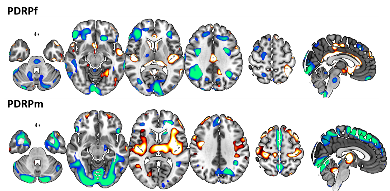
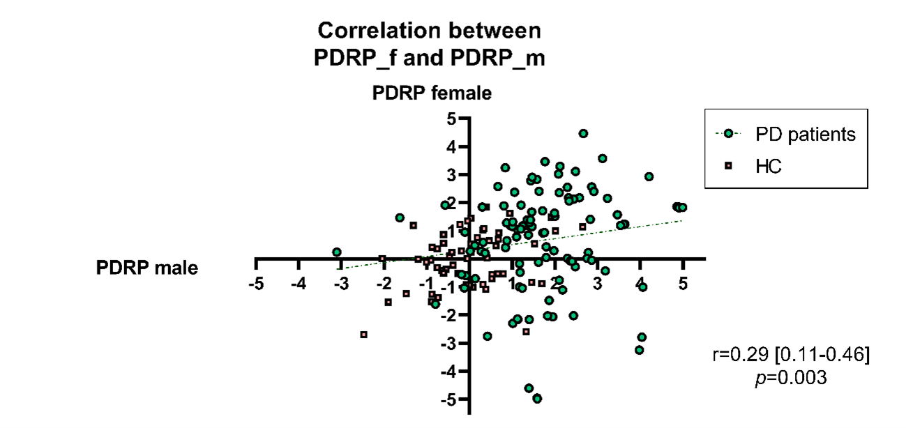
Method: We conducted a multicenter retrospective cohort study that included a total of 144 patients with a diagnosis of PD and 75 Healthy Controls (HC). Each patient had been referred for FDG-PET imaging in the first decade of the disease course. To assess brain metabolic gender differences, we used both a univariate Statistical Parametric Mapping (SPM) procedure and a multivariate Scaled Subprofile Model/Principal Component Analysis (SSM/PCA). In the SSM/PCA approach, we selected two identification cohorts, one composed of PD and HC males and the other composed of PD and HC females. Then, we obtained a PDRP-male and PDRP-female. To compare the patterns we use three approaches. First, we performed a voxel correlation between patterns. Second, we computed the individual scores of identified patterns and performed a correlation test of individual scores from both patterns. Finally, we visually analyzed both
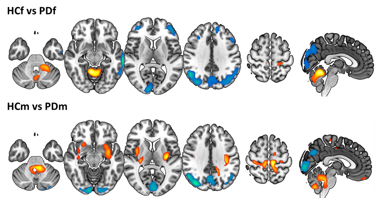
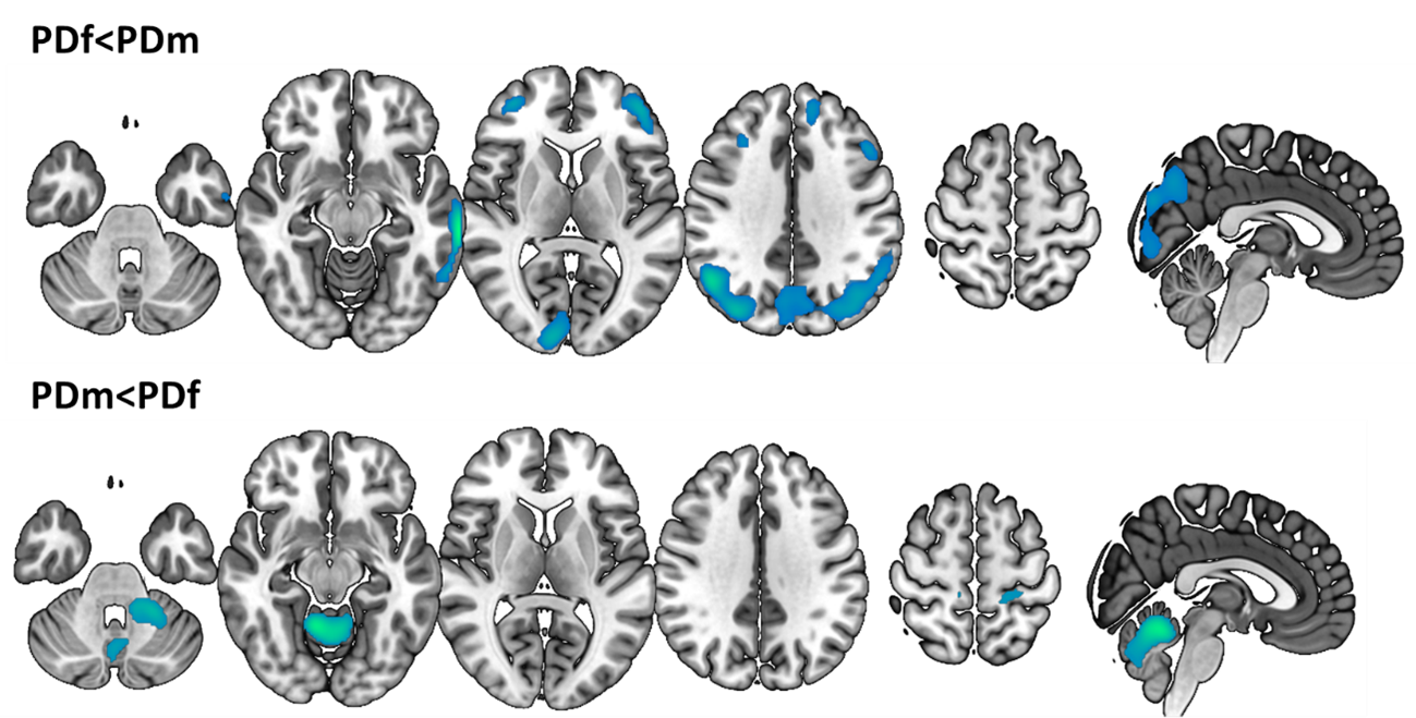
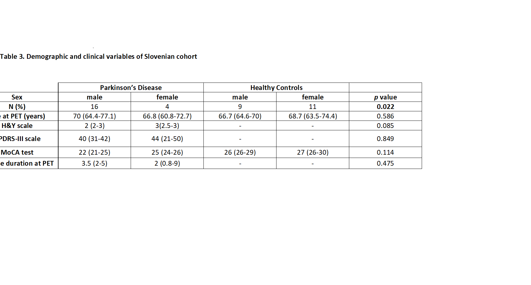
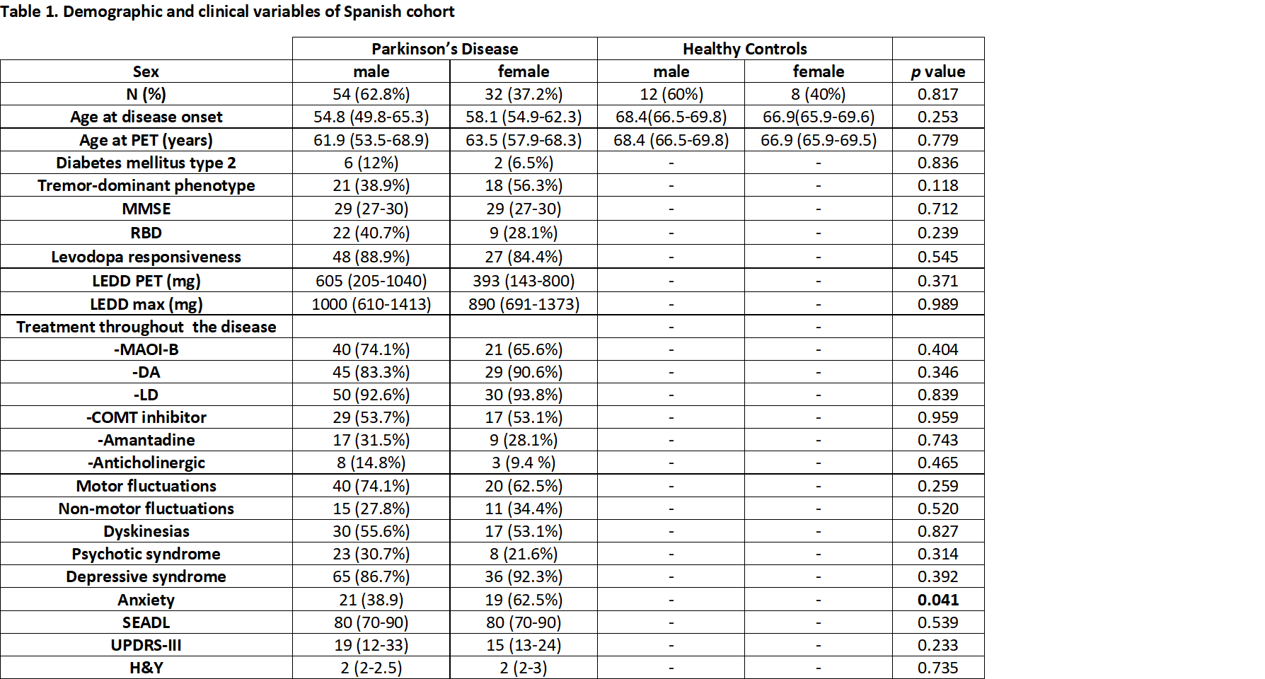
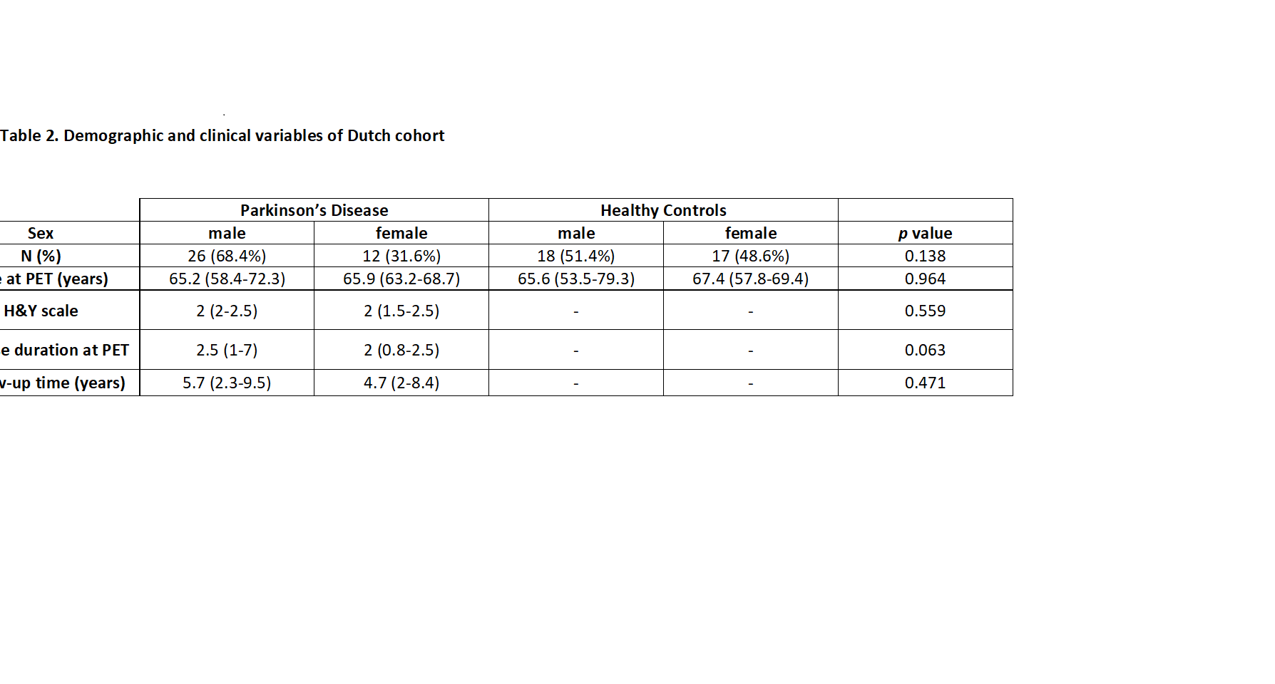
Results: We included three cohorts. Spanish and Dutch cohorts showed similar age and sex distribution among groups [Table 1-2]. Slovenian cohort showed similar age, but more proportion of males in PD group [Table 3]. In the Spanish cohort, female PD patients showed more anxiety and lower scores in previously identified global PDRP [Table 1]. Based on SPM t-maps, PD males and females showed hypermetabolism in the cerebellum and primary sensory-motor cortex and hypometabolism in parieto-occipital cortex. PD females showed an additional hypometabolism in the prefrontal cortex and PD males hypermetabolism in the striatum [Figure 1]. On the other hand, PD gender-related metabolic patterns are illustrated in [Figure 2]. Voxel-correlation between patterns was statistically significant but weak (r=0.152, p<0.001). The correlation between individual scores was statistically significant but weak [Figure 3]
Conclusion: Brain metabolic abnormalities in PD patients may be influenced by gender. Despite similar regions involved in males and females in PD patients, there are also regional differences that might be related to clinical profiles
References: [1] Arabia G, De Martino A, Moro E. Sex and gender differences in movement disorders: Parkinson’s disease, essential tremor, dystonia and chorea [Internet]. 1st ed. Vol. 164, International Review of Neurobiology. Elsevier Inc.; 2022. 101–128 p. Available from: http://dx.doi.org/10.1016/bs.irn.2022.06.010
[2] Martí-Andrés, G., et al. Clinical correlates of Parkinson’s disease metabolic pattern: not only a diagnostic tool. 25th international congress of Parkinson ́s disease and movement disorders. MDS virtual congress 2020.
To cite this abstract in AMA style:
G. Martí-Andrés, E. Prieto-Azcarate, CA. Espinoza-Vinces, M. Riverol, S. Meles, KL. Leenders, P. Tomse, M. Perovnik, M. Trost, J. Arbizu, MR. Luquin-Puido. Clinical and radiological gender differences in Parkinson’s disease [abstract]. Mov Disord. 2023; 38 (suppl 1). https://www.mdsabstracts.org/abstract/clinical-and-radiological-gender-differences-in-parkinsons-disease/. Accessed January 2, 2026.« Back to 2023 International Congress
MDS Abstracts - https://www.mdsabstracts.org/abstract/clinical-and-radiological-gender-differences-in-parkinsons-disease/

