Category: Myoclonus
Objective: To report spread of myoclonus in Subacute Sclerosing Pan Encephalitis (SSPE)
Background: SSPE is a slowly progressive encephalitis caused by mutant measles virus with mutations in M gene. SSPE is clinically characterized by behavioral abnormalities, cognitive decline, periodic myoclonus and vision abnormalities. The progression of myoclonus in SSPE has been poorly characterized.
Method: A 17-year-old male presented with diminution of vision for 6 months, abnormal jerky movements of proximal upper limbs and lower limbs for 4 months, behavioral changes in the form of memory disturbances, repeating phrases and bladder bowel incontinence for 4 months.Examination revealed mutism, generalized rigidity with slow pseudo periodic myoclonus involving bilateral upper and lower limbs. There was a characteristic phenomenon of left superior gaze deviation (slow myoclonus) for a brief period following few milliseconds after the myoclonus of the limbs.
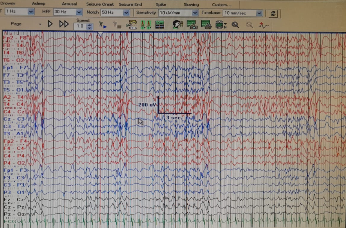
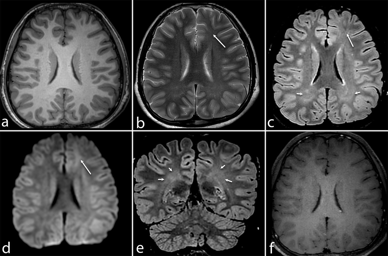
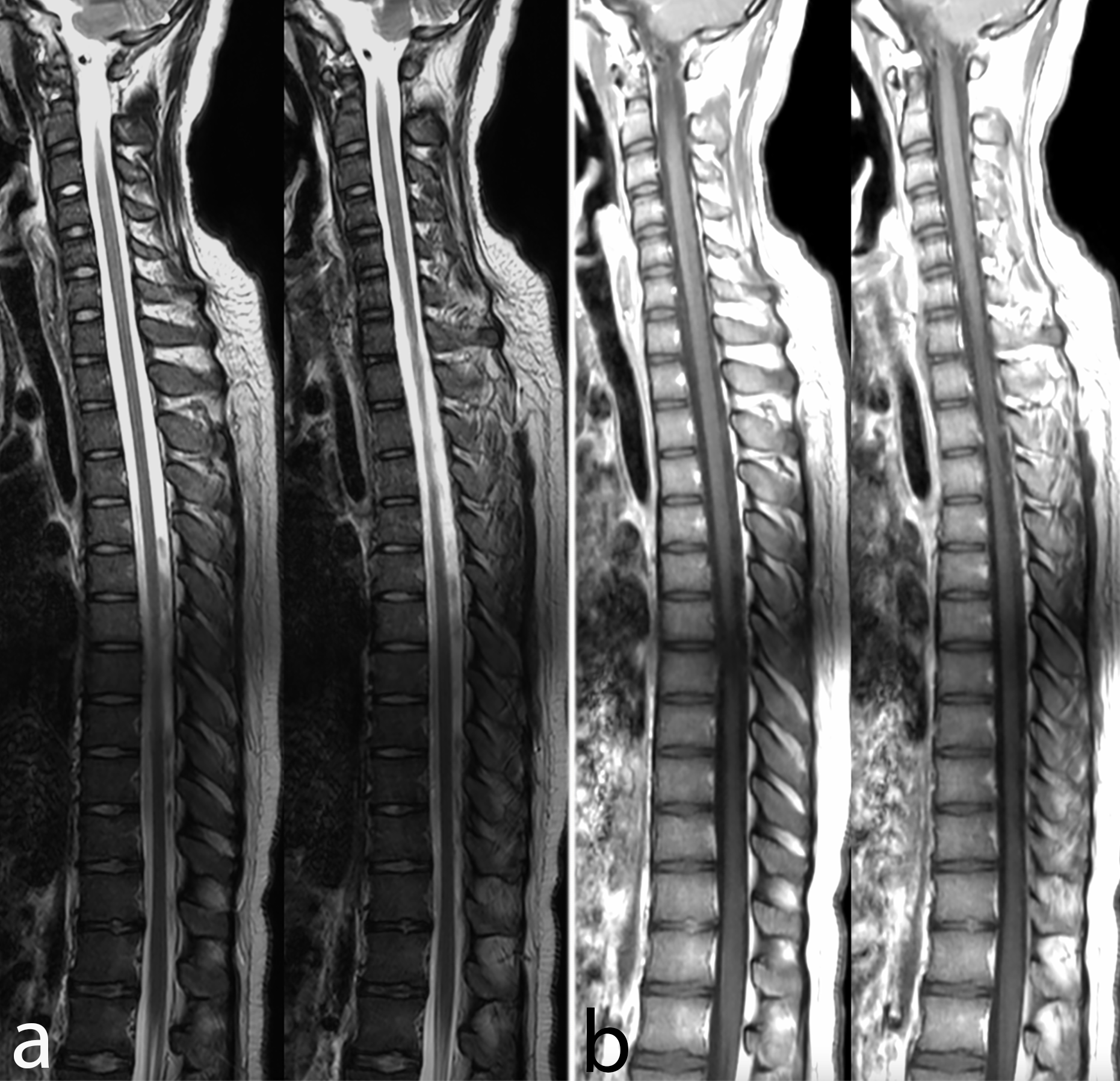
Results: Electroencephalography revealed a periodic burst suppression pattern with background slowing. Bursts were in the form of 2-2.5 Hz, 90-110 millivolts, sharp and wave discharges with inter-periodic interval of 3-4 seconds (Figure 1). Magnetic resonance imaging of the brain revealed hyperintensities in bilateral frontal subcortical areas with normal brain-stem and spinal cord (Figure 2, 3).CSF/Serum quotient was positive at 4.55 (with normal being <1.3).Our patient fell in Stage 3 of Jabbour clinical staging and IIIB of Gascon stage.1,2 His poly-electromyography recording showed an initial onset of myoclonus in right tibialis anterior (TA) followed by right sternocleidomastoid (SCM). Right levator palpebrae superioris (LPS) did show electromyography recording about one second following the recording in right SCM.The burst durations of SCM were about 100 milli-secs while those of TA of 50 milli-sec and LPS of 30 milli-sec (Figure 4).This is in line with a recent study where myoclonus in SSPE have been shown to involve brain stem at a later clinical stage.3 However, initial triggering in tibial muscles and subsequent slow spread to eyeballs from brain stem have not been described.
Conclusion: Our findings of poly-electromyography may suggest brain stem myoclonus (as depicted by SCM recording with slow progression to eyeballs) may be triggered or preceded by a segmental peripheral myoclonus affecting tibial group of muscles.
References: 1. Brismar J, Gascon GG, von Steyern KV, Bohlega S. Subacute sclerosing panencephalitis: evaluation with CT and MR. AJNR Am J Neuroradiol. 1996 Apr;17(4):761-72.
2. Garg RK. Subacute sclerosing panencephalitis. Postgrad Med J. 2002 Feb;78(916):63-70.
3. Ser MH, Gündüz A, Demirbilek V, Yalçınkaya C, Nalbantoğlu M, Coşkun T, Kızıltan M. Progression of myoclonus subtypes in subacute sclerosing panencephalitis. Neurophysiol Clin. 2021 Dec;51(6):533-540. doi: 10.1016/j.neucli.2021.07.001.
To cite this abstract in AMA style:
A. Das, J. George, A. Garg, A. Srivastava. Initial triggering in tibial group and subsequent slow spread of myoclonus from brain stem to eye-balls in Subacute Sclerosing Pan Encephalitis [abstract]. Mov Disord. 2023; 38 (suppl 1). https://www.mdsabstracts.org/abstract/initial-triggering-in-tibial-group-and-subsequent-slow-spread-of-myoclonus-from-brain-stem-to-eye-balls-in-subacute-sclerosing-pan-encephalitis/. Accessed March 31, 2025.« Back to 2023 International Congress
MDS Abstracts - https://www.mdsabstracts.org/abstract/initial-triggering-in-tibial-group-and-subsequent-slow-spread-of-myoclonus-from-brain-stem-to-eye-balls-in-subacute-sclerosing-pan-encephalitis/


