Category: Tremor
Objective: We investigated reconstructing the entire dentato-rubro-thalamic tract (DRT) by an anatomical “rubral wing”-based tractography and compare its correlation with clinical outcomes to those of a deterministic tractography method of Bot et al.
Background: The DRT appears to be an important structure for identifying when planning MRI-guided high-intensity focused ultrasound (HIFU) thalamotomy for tremor. The rubral wing (RW) of the DRT can be visualized directly using the fast gray matter acquisition T1 inversion recovery (FGATIR)
Method: We retrospectively reviewed the data of 23 patients with tremor that underwent HIFU thalamotomy. DRT tractography, ipsilateral to the site of lesion was separately created by 2 neurosurgeons, based on either the traditional method of Bot et al. (Bot-DRT) or the anatomical “rubral-wing” method (RW-DRT). The anatomic method was based on ROIs of the rubral wing and the SCP, as depicted on the FGATIR sequence. Dice coefficient was used to measure differences between the 2 tractography methods and inter-observer reproducibility for each method. Post-HIFU lesion in reference to each of the tractography methods was correlated with tremor outcomes and side effects.
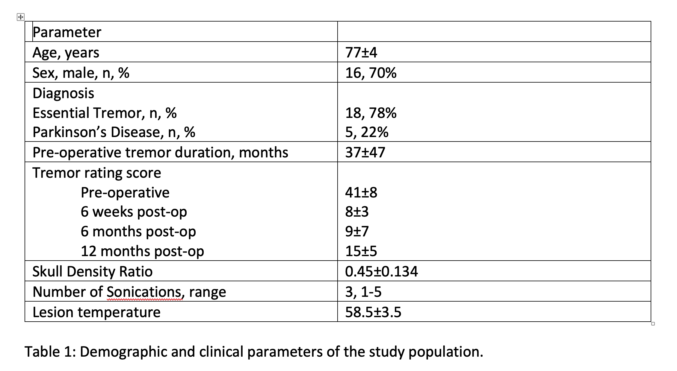
Results: Good tremor control was achieved in 21/23 patients (91%) 6 weeks. Side effects were evident in 14/23 patients (60%) in 6 weeks, mainly as temporary mild imbalance.
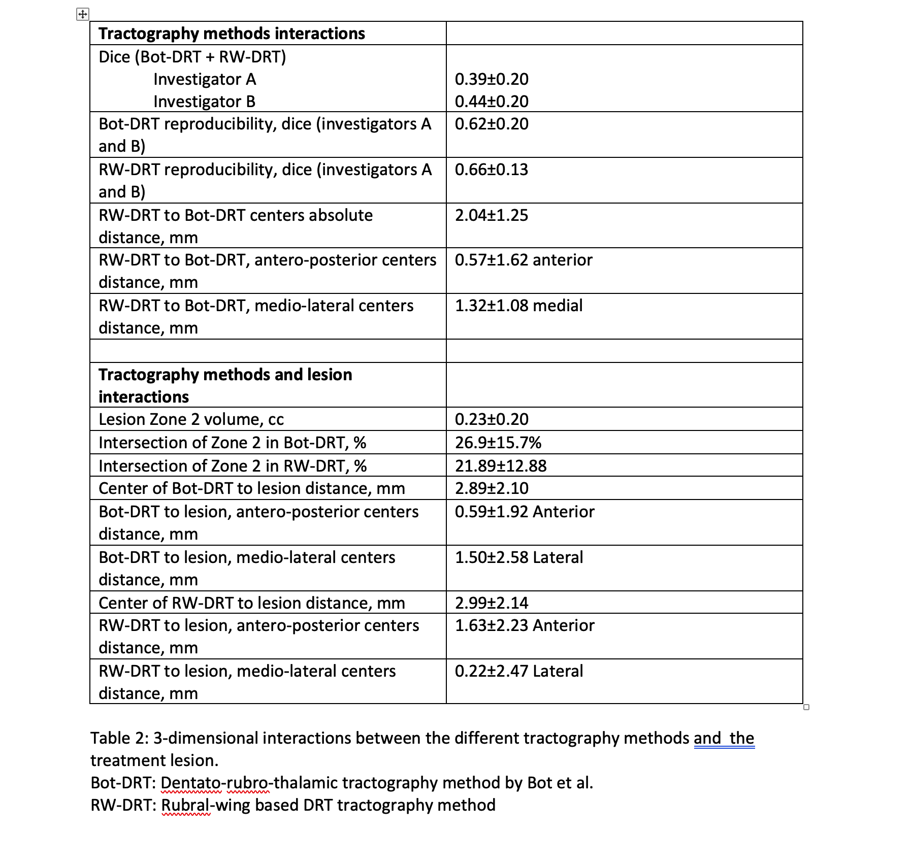
Bot-DRT involved the rubral wing and RW-DRT intersected with all 4 ROIs of Bot-DRT in all 23 cases. However, both investigator A and B found low correlation indices between the Bot and RW tractography methods, with DICE coefficients of 0.39±0.20 and 0.44±0.20, respectively, p=0.15. There were high reproducibility indices between the 2 investigating neurosurgeons in depicting both the Bot and RW methods with DICE coefficients of 0.62±0.20 and 0.66±0.13, respectively, p=0.40.
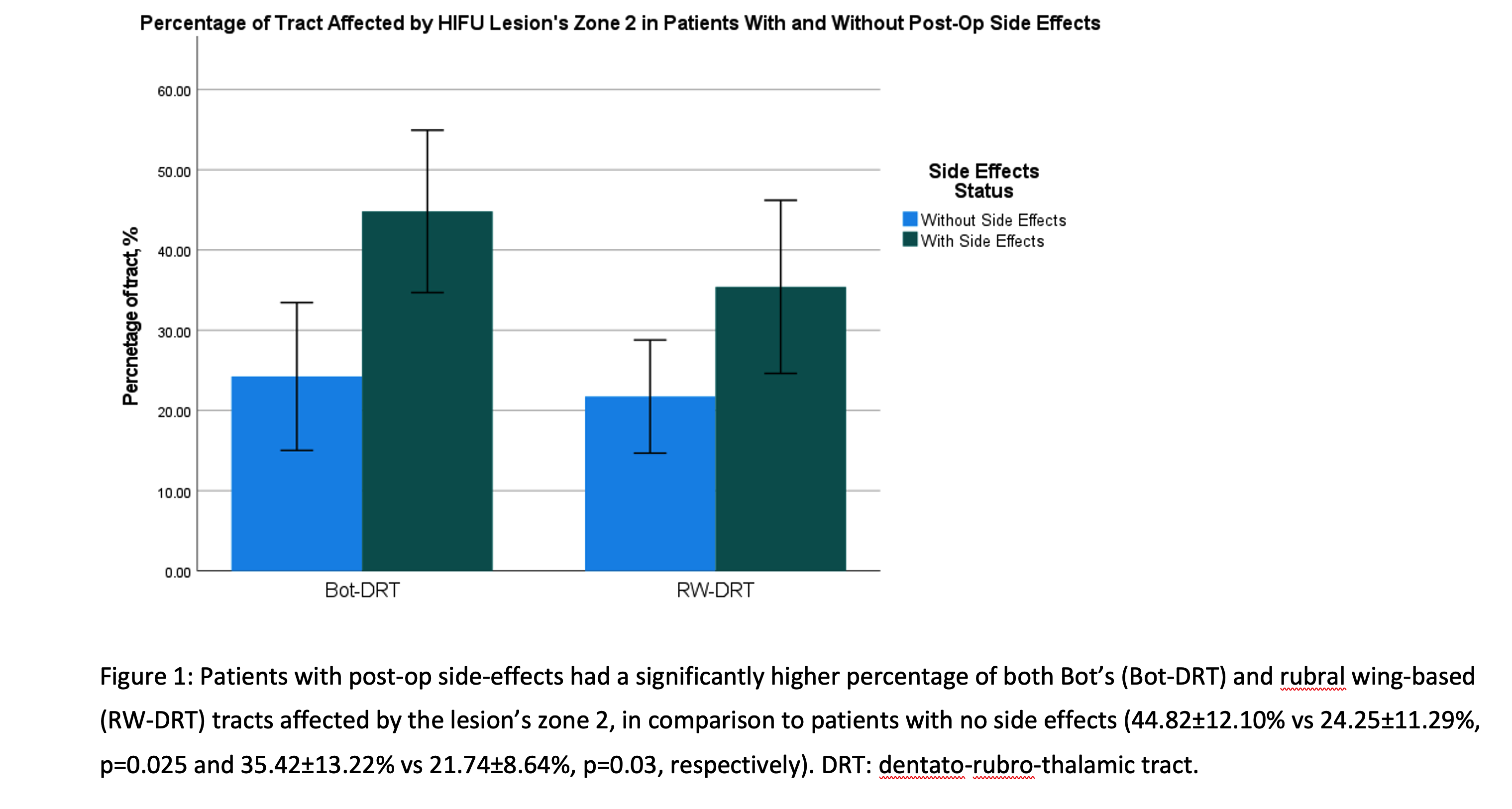
Among those with good tremor control 6-months post-op (n=12), patients with side effects (n=6) had a significantly higher percentage of both Bot-DRT and RW-DRT tracts affected by the lesion, (44.82±12.10% vs 24.25±11.29%, p=0.025 and 35.42±13.22% vs 21.74±8.64%, p=0.03, respectively).
Conclusion: Tractography of the DRT can be simply reconstructed by direct anatomical visualization of the rubral wing. This tractography shares similarities with an older method suggested by Bot, but its fibers pass antero-medially to it.
To cite this abstract in AMA style:
A. Berger, J. Chung, Z. Schnurman, L. Pan, T. Shepherd, A. Mogilner. Reconstruction of the dentato-rubro-thalamic tract based on the anatomy of the rubral wing [abstract]. Mov Disord. 2023; 38 (suppl 1). https://www.mdsabstracts.org/abstract/reconstruction-of-the-dentato-rubro-thalamic-tract-based-on-the-anatomy-of-the-rubral-wing/. Accessed December 19, 2025.« Back to 2023 International Congress
MDS Abstracts - https://www.mdsabstracts.org/abstract/reconstruction-of-the-dentato-rubro-thalamic-tract-based-on-the-anatomy-of-the-rubral-wing/


