Category: Dystonia: Pathophysiology, Imaging
Objective: To access striatal matrix and striosome compartments in patients with idiopathic upper limb dystonia using diffusion tensor imaging
Background: The striatum is an essential hub in the motor system associated with dystonia and other movement disorders. The function of the striosomes and matrix in motor control is not clear. A recently developed method using diffusion tensor imaging (DTI) enables us to distinguish compartments of the striatum, namely matrixes-like and striosomes-like voxels, in vivo. Evaluating the striatum in terms of matrixes and striosomes may allow the understanding of the neurobiology of dystonia and ultimately lead to more targeted treatments.
Method: We analyzed 3T MRI images from 26 patients with idiopathic upper limb dystonia aged 43.88 ± 11.32 years (SD; range 19–60) with a mean disease duration of 12.55 ± 10.25 years (SD; range 1–25) and healthy controls aged 39.42 ± 11.42 years (SD; range 19–58).The striatum was parcellated by targeting cortical regions that favored striosomes (pregenual anterior cingulate, posterior orbitofrontal, anterior insular cortex, and basolateral amygdala) and matrix-favoring areas (gyrus rectus, supplementary motor area, and primary sensory and motor cortex). The bilateral striatum was assessed for changes in volume and mean fractional anisotropy (FA) value.
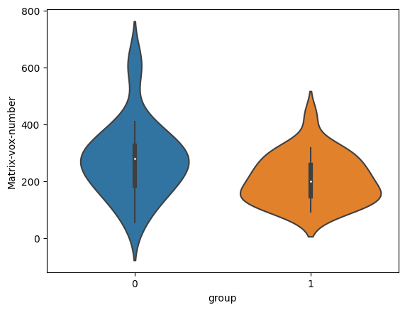
Results: Patients show significant reductions of left Matrix-like voxels volume relative to controls (p= 0.022), with a moderate effect size (Cohen’s d = 0.640)[Figure 1]. No difference was observed in the right striatum compartments.
Conclusion: By parcellating the striatum into striosome and matrix-like voxels, we showed that patients with idiopathic dystonia have a reduced volume in matrix-like voxels in agreement with anatomopathological findings of some genetic types of dystonia. Even in non-degenerative dystonias, volume differences may reflect an imbalance between matrix and striosome signaling, ultimately favoring the direct pathway.
References: Waugh, J. L., Hassan, A. A., Kuster, J. K., Levenstein, J. M., Warfield, S. K., Makris, N., … & Blood, A. J. (2022). An MRI method for parcellating the human striatum into matrix and striosome compartments in vivo. Neuroimage, 246, 118714.
To cite this abstract in AMA style:
AJM. Paulo, J. Waugh, JPQ. de Paiva, JR. Sato, DD. de Faria, V. Borges, SMA. Silva, HB. Ferraz, PMC. Aguiar. In vivo assessment of striatal compartments in patients with idiopathic upper limb dystonia [abstract]. Mov Disord. 2023; 38 (suppl 1). https://www.mdsabstracts.org/abstract/in-vivo-assessment-of-striatal-compartments-in-patients-with-idiopathic-upper-limb-dystonia/. Accessed February 23, 2026.« Back to 2023 International Congress
MDS Abstracts - https://www.mdsabstracts.org/abstract/in-vivo-assessment-of-striatal-compartments-in-patients-with-idiopathic-upper-limb-dystonia/

