Category: Parkinsonism, Atypical: PSP, CBD
Objective: To compare brain MRI morphometry and regional hypometabolism among the clinical predominance-types of Progressive Supranuclear Palsy.
Background: Recently, MDS-PSP criteria for the clinical diagnosis of PSP and 8 predominance-types were proposed.1 MRI morphometry and regional hypometabolism in the newly classified predominance-types1 is less known.
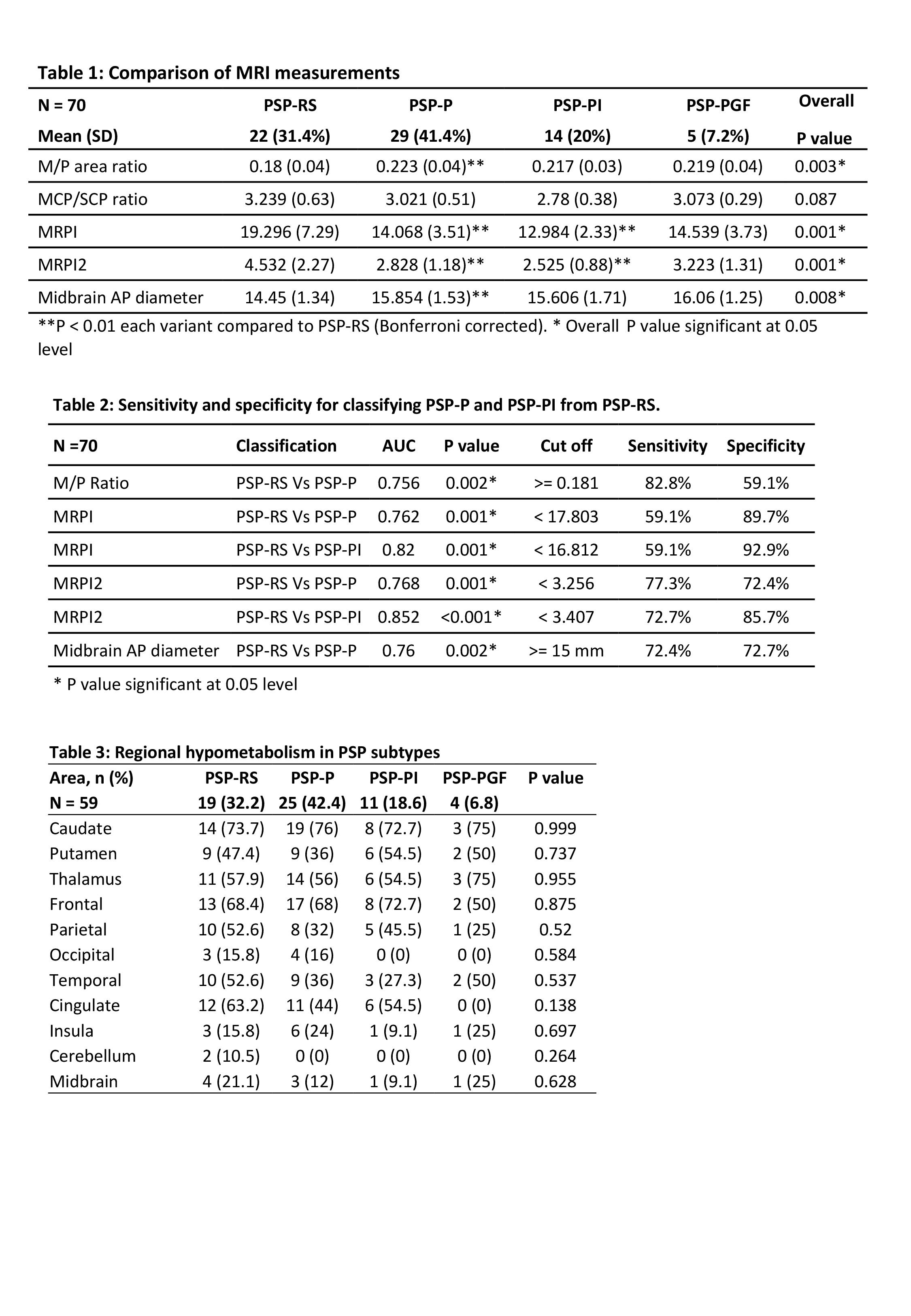
Method: All cases of PSP diagnosed and categorised using the MDS-PSP clinical criteria,1,2 were enrolled prospectively. The MRI measures were ratios of midbrain/ pons (M/P) area and middle cerebellar peduncle/ superior cerebellar peduncle width (MCP/ SCP), MR Parkinsonism Indices (MRPI and MRPI 2) and midbrain antero-posterior diameter, done by 2 blinded MR imaging specialists. A blinded, experienced molecular imaging specialist rated the 18FFDG PET images in pre-defined regions as normal, hypo or hypermetabolic, and corroborated it by software-based quantitative analysis, matching images with age and gender -matched controls. Fisher’s exact test and One-way ANOVA test were used to compare variables and post-hoc analysis was done with Bonferroni correction for multiple comparisons. Pearson’s correlation was used to test associations. The ROC curve coordinate point with highest Youden index was considered the optimal cut-off value.
Results: 80 cases of PSP completed the study. There were only 2 PSP-CBD and 1 speech/language types which were not included. 70 MRI and 59 PET scans were completed. Some participants consented only for one of the 2 tests (both tests in 59 cases). Inter-rater correlations were very good or excellent. M/P area ratio and midbrain AP diameter were lower in PSP-RS than PSP-P while MRPI and MPRI 2 were higher in PSP RS compared to PSP-P and PSP-PI (Table 1 and 2). Hypometabolic regions did not differ among the PSP types (Table 3). There were small, but significant (P<0.05) correlations between both MRPI and MRP2 with scores of PSP rating scale (r=0.4&0.35), UPDRSIII (r=0.39&0.34), MOCA (r=-0.39&-0.36) and FAB (r=-0.47&-0.4).
Conclusion: MRI can differentiate PSP-RS from both PSP-P and PSP-PI, with the highest specificity for MRPI. Regional hypometabolism is similar among the types of PSP, irrespective of the clinical predominance of the core features.
References: 1. Höglinger GU, Respondek G, Stamelou M et al. Clinical diagnosis of progressive supranuclear palsy: The movement disorder society criteria. Mov Disord. 2017 Jun;32(6):853-864.
2. Grimm MJ, Respondek G, Stamelou M, et al. How to apply the movement disorder society criteria for diagnosis of progressive supranuclear palsy. Mov Disord 2019;34(8):1228-1232.
To cite this abstract in AMA style:
H. Chovatiya, K. Pillai, R. Kamble, S. Santhamma, P. Rajeswari, M. Chacko, A. Kishore. MRI morphometry and regional hypometabolism in the clinical predominance-types of Progressive Supranuclear Palsy [abstract]. Mov Disord. 2023; 38 (suppl 1). https://www.mdsabstracts.org/abstract/mri-morphometry-and-regional-hypometabolism-in-the-clinical-predominance-types-of-progressive-supranuclear-palsy/. Accessed January 15, 2026.« Back to 2023 International Congress
MDS Abstracts - https://www.mdsabstracts.org/abstract/mri-morphometry-and-regional-hypometabolism-in-the-clinical-predominance-types-of-progressive-supranuclear-palsy/

