Category: Dystonia: Pathophysiology, Imaging
Objective: To compare key white matter motor pathways between those diagnosed with dystonia and unaffected controls, using data collected by the UK Biobank, processed and analysed using highly optimised techniques.
Background: Multimodal imaging, animal model and human histopathological work have implicated network dysfunction in dystonia pathogenesis, with the cerebellum, basal ganglia and sensorimotor cortex, and their connecting pathways, seemingly key. Diffusion MRI (dMRI) findings in dystonia cohorts are mixed, although changes to cerebellar white matter projections, and white matter deep to the primary sensory-motor cortices are consistently highlighted. The UK Biobank includes data for >500,000 participants, with dMRI data for a proportion of individuals (n=42,565). While standardised imaging processing pipelines exist, optimised methodology is essential in studies of movement disorder cohorts to account for movement related artifacts.
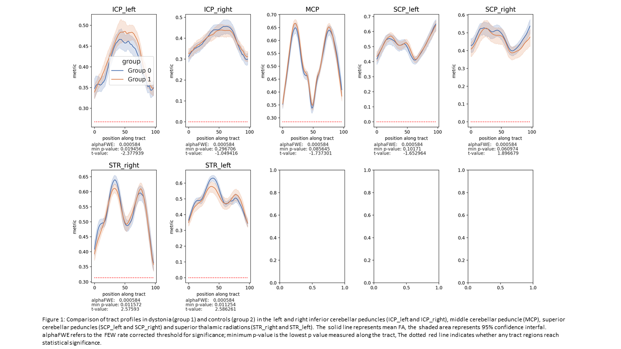
Method: The dystonia cohort was identified using hospital (ICD-10) and primary care (Read codes) data[1], and an age and gender matched control cohort was derived. dMRI data was preprocessed using a bespoke pipeline including correction for Gibbs ringing, signal drift, subject motion, and outliers[2]. dMRI-derived measures were profiled along reconstructed tracts (tractometry)[3], assessing FA (fractional anisotropy) along the cerebellar peduncles and superior thalamic radiations, with family-wise error multiple comparison correction.
Results: Seventy-eight individuals within the dystonia cohort had undergone DTI, along with a control cohort of 773. A subset (55 dystonia cases, 40 controls) was included in this interim analysis. Comparison of FA via tractometry of the cerebellar peduncles and superior thalamic radiations identified no significant differences between the dystonia cohort and controls[figure1].
Conclusion: The cerebellar outflow and thalamo-cortical white matter differences previously indicated were not evident in this cohort. This may be due to the multiple dystonia subtypes combined within this group, the potential for a lack of white matter involvement in these regions or the poor biological specificity of FA as a measure, resulting in poor sensitivity to subtle tract differences. More detailed work with application of more biologically specific measures, together with evaluation of individual dystonia subtypes is needed in this cohort.
References: [1] Bailey GA, Rawlings A, Torabi F, Pickrell O, Peall KJ. Adult-onset idiopathic dystonia: A national data-linkage study to determine epidemiological, social deprivation, and mortality characteristics. Eur J Neurol. 2022;29(1):91-104.
[2] Tax CMW, Bastiani M, Veraart J, Garyfallidis E, Okan Irfanoglu M. What’s new and what’s next in diffusion MRI preprocessing. Neuroimage. 2022;249:118830.
[3] Wasserthal J, Neher P, Maier-Hein KH. TractSeg – Fast and accurate white matter tract segmentation. NeuroImage. 2018;183:239-53.
To cite this abstract in AMA style:
C. Maciver, M. Wadon, G. Bailey, A. Schalkamp, C. Sandor, D. Jones, C. Tax, K. Peall. Diffusion MRI tractometry findings in Dystonia- A UK Biobank study [abstract]. Mov Disord. 2022; 37 (suppl 2). https://www.mdsabstracts.org/abstract/diffusion-mri-tractometry-findings-in-dystonia-a-uk-biobank-study/. Accessed February 2, 2026.« Back to 2022 International Congress
MDS Abstracts - https://www.mdsabstracts.org/abstract/diffusion-mri-tractometry-findings-in-dystonia-a-uk-biobank-study/

