Objective: To investigated the integrity of white matter tracts within basal ganglia-frontal networks in PD patients with and without apathy, and relate these to specific changes in cognitive processes underlying goal directed behavior.
Background: Apathy is one of the most common neuropsychiatric symptoms, and a major contributor to the disability and decreased quality of life of patients with Parkinson’s Disease (PD) and other neurodegenerative diseases. It is likely that apathy results from the disruption of a network of brain regions implicated in normal goal directed behavior. Effort Based Decision Making (EBDM) paradigms are a valid approach to neuroanatomically dissociate effort and reward components underlying motivational processes, and secondarily apathetic behaviors.
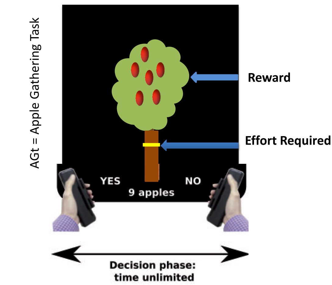
Method: Multi-shell diffusion MRI has so far been obtained in 9 PD participants, out of a planned n=100. Apathy was measured by the Starkstein Apathy Scale (SAS). A fiber-based analysis (FBA) was performed using MRtrix3 and a summary metric of white matter integrity called fiber and cross-section density (FDC) was obtained. All patients are undertaking an EBDM paradigm in which they decide whether it is worth exerting physical effort for reward (the Apple Gathering task [AGt] – Fig 1). Computational analysis using a Hierarchical Drift diffusion Model allows dissociation of reward and effort effects on choice and decision time, creating a ‘fingerprint’ of an individual’s motivational processes that can be related to neuroimaging and traditional apathy assessment methods.
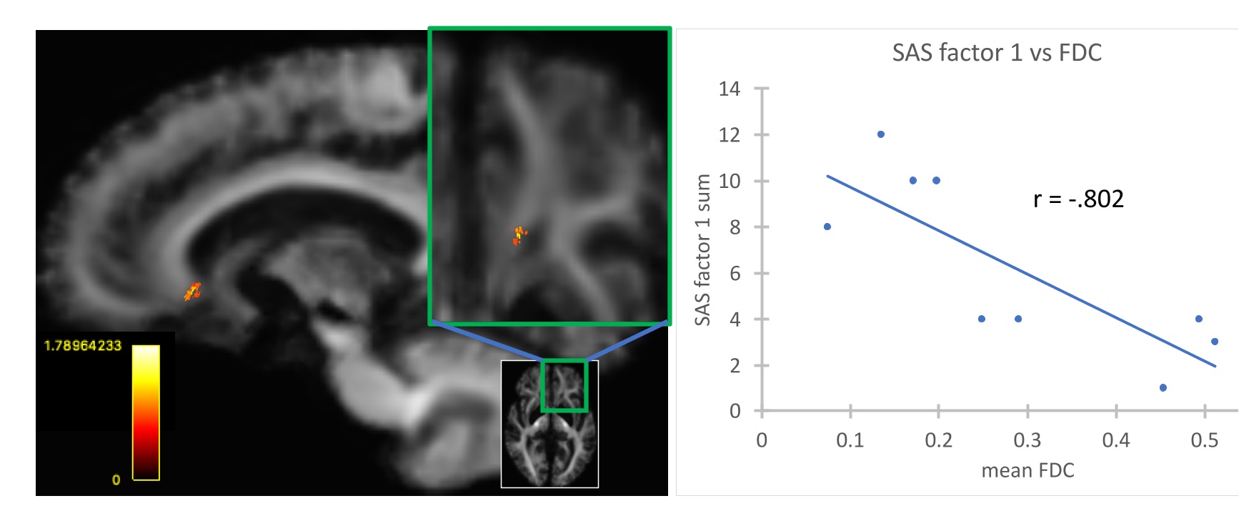
Results: In 9 PD subjects (age = 56 ± 9 years, 3 females/6 males, SAS = 11.3 ± 6.04 with 3 apathetic/6 non-apathetic), a correlation analysis between apathy severity and FDC has been conducted. Apathy severity was negatively correlated with FDC in the left corona radiata/forceps minor (connecting frontal-basal ganglia structures) with a maximum effect size (Cohen’s D) of 0.79. Fig. 2. Imaging results of the expanded cohort and relationships to reward and effort processing will be presented.
Conclusion: White matter atrophy may contribute to apathy features exhibited in PD and other neurodegenerative processes. The location of the damage may be specific to each disease but ultimately affect the same motivation related network. Additional results characterizing cognitive motivational processes in relation to neuroimaging measures, EBDM and real-world behaviors in PD will be presented.
References: Prange, S. et al. Early limbic microstructural alterations in apathy and depression in de novo Parkinson’s disease. Movement disorders : official journal of the Movement Disorder Society 34, 1644- 1654, doi:10.1002/mds.27793 (2019).
Sun, H. H. et al. Alterations of regional homogeneity in Parkinson’s disease with “pure” apathy: A resting-state fMRI study. Journal of affective disorders 274, 792-798, doi:10.1016/j.jad.2020.05.145 (2020).
Le Heron, C. et al. Distinct effects of apathy and dopamine on effort-based decision-making in Parkinson’s disease. Brain : a journal of neurology 141, 1455-1469, doi:10.1093/brain/awy110 (2018).
Tournier, J. D. et al. MRtrix3: A fast, flexible and open software framework for medical image processing and visualisation. NeuroImage 202, 116137, doi:10.1016/j.neuroimage.2019.116137 (2019).
To cite this abstract in AMA style:
C. Le Heron, E. Huey, N. Vanegas-Arroyave, Y. Gazes, S. Lee. Neuroanatomical underpinnings of apathy in Parkinson’s Disease [abstract]. Mov Disord. 2022; 37 (suppl 2). https://www.mdsabstracts.org/abstract/neuroanatomical-underpinnings-of-apathy-in-parkinsons-disease/. Accessed December 19, 2025.« Back to 2022 International Congress
MDS Abstracts - https://www.mdsabstracts.org/abstract/neuroanatomical-underpinnings-of-apathy-in-parkinsons-disease/


