Category: Neuroimaging (Non-PD)
Objective: To better understand the role of FDG-PET for identifying dementia with Lewy bodies (DLB) in the context of Alzheimer’s disease (AD)-like amnestic presentations, we analysed individual FDG-PET patterns in a large cohort of patients clinically diagnosed with AD dementia (ADD) or amnestic MCI (aMCI), identifying those with FDG-PET features suggestive of DLB. We provide an in-depth characterization of this subgroup with respect to pathological and clinical features.
Background: Clinical misdiagnosis is common between AD and DLB [1]. In FDG-PET, a pattern of pronounced posterior-occipital hypometabolism with relative sparing of the posterior cingulate cortex and the medial temporal lobe is a well-established imaging signature suggestive of DLB [2,3]. The utility of this imaging signature in the absence of DLB specific symptomatology is yet to be explored.
Method: Our study included 1214 patients from the Alzheimer’s Disease Neuroimaging Initiative (aMCI=909, ADD=305). Baseline FDG-PET scans were classified as suggestive of AD (sAD) or DLB (sDLB) by using a logistic regression classifier previously trained on a separate set of autopsy-confirmed AD and DLB cases. sAD and sDLB subgroups were compared on Aβ- and tau-PET imaging characteristics. We also compared longitudinal domain-specific cognitive performance (memory and executive function composite scores) and the risk of developing hallucinations on follow-up (3y for ADD, 6y for aMCI).
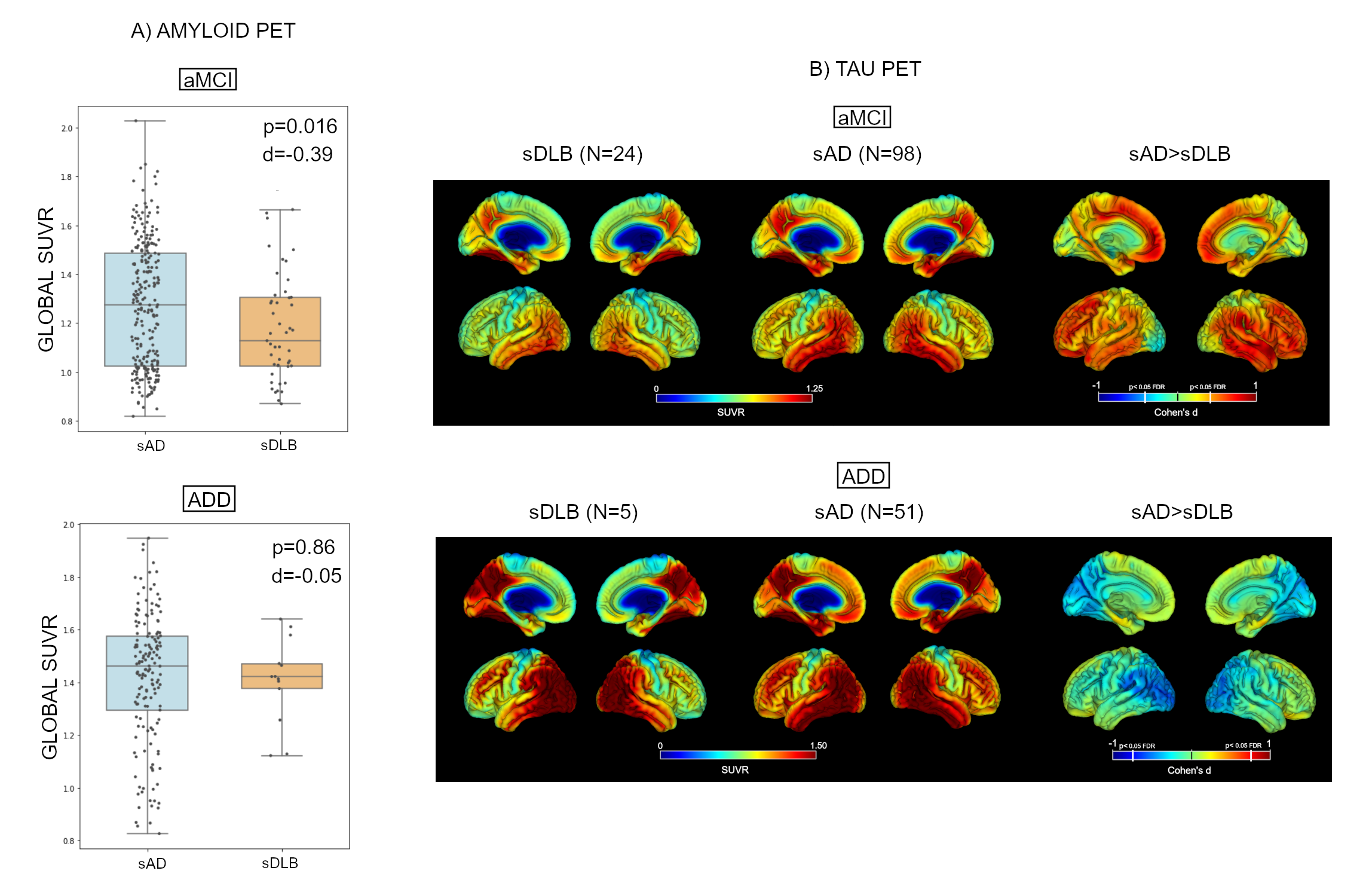
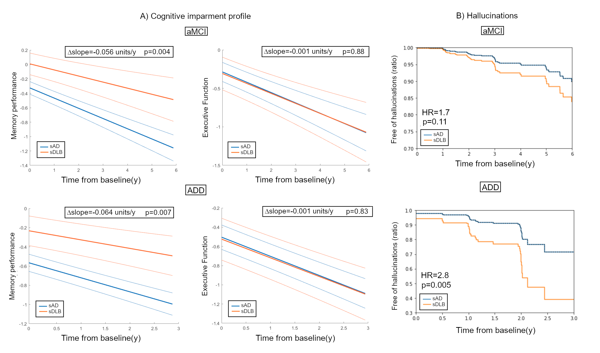
Results: Of 414 aMCI and 259 ADD patients that showed evidence of regional hypometabolism in AD- or DLB-relevant areas, 69 aMCI (17%) and 28 ADD (11%) patients were classified as sDLB. sDLB aMCI patients showed significantly lower Aβ PET and tau PET burden than sAD (Fig. 1), but these differences were not observed for ADD patients. sDLB patients showed better memory performance at baseline and less memory decline (aMCI: p=0.004; AAD: p=0.007) compared to sAD, but comparable executive function (Fig. 2A). sDLB patients had a higher risk of developing hallucinations, though this did only reach significance for ADD (p>0.005, HR=2.8; aMCI: p=0.11, HR=1.7) (Fig. 2B).
Conclusion: FDG-PET signatures suggestive of DLB are present in a sizeable subset of clinically-diagnosed ADD and aMCI patients. These patients showed reduced AD pathology and a distinctive clinical evolution.
References: [1] AJ Thomas et al. “Improving the identification of dementia with Lewy bodies in the context of Alzheimer’s-type dementia” (Alzheimer’s Res Ther. 2018). DOI: 10.1186/s13195-018-0356-0
[2] K Kantarci et al. “FDG PET metabolic signatures distinguishing prodromal DLB and prodromal AD” (Neuroimage Clin. 2021). DOI: 10.1016/j.nicl.2021.102754
[3] Graff-Radford et al. “18 F-fluorodeoxyglucose positron emission tomography in dementia with Lewy bodies” (Brain Commun. 2020). DOI: 10.1093/braincomms/fcaa040
To cite this abstract in AMA style:
J. Silva-Rodríguez, MA. Labrador-Espinosa, A. Moscoso, M. Schöll, P. Mir, M. Grothe. FDG-PET features of Lewy body dementia among patients with amnestic presentations typical for Alzheimer’s disease [abstract]. Mov Disord. 2022; 37 (suppl 2). https://www.mdsabstracts.org/abstract/fdg-pet-features-of-lewy-body-dementia-among-patients-with-amnestic-presentations-typical-for-alzheimers-disease/. Accessed October 18, 2025.« Back to 2022 International Congress
MDS Abstracts - https://www.mdsabstracts.org/abstract/fdg-pet-features-of-lewy-body-dementia-among-patients-with-amnestic-presentations-typical-for-alzheimers-disease/


