Category: Parkinson's Disease: Neuroimaging
Objective: We aimed to characterize the dynamic functional connectivity (DFC) in people with FOG based on high temporal-resolution functional MRI (fMRI).
Background: Freezing of gait (FOG) is a gait disorder that often accompanies Parkinson’s disease (PD) and is closely associated with abnormalities in the function of brain networks[1]. The current understanding of brain functional organization in FOG was built on the assumption that the functional connectivity (FC) of brain networks is static, but these change dynamically over time[2]. The characteristics of DFC in FOG is still unclear.
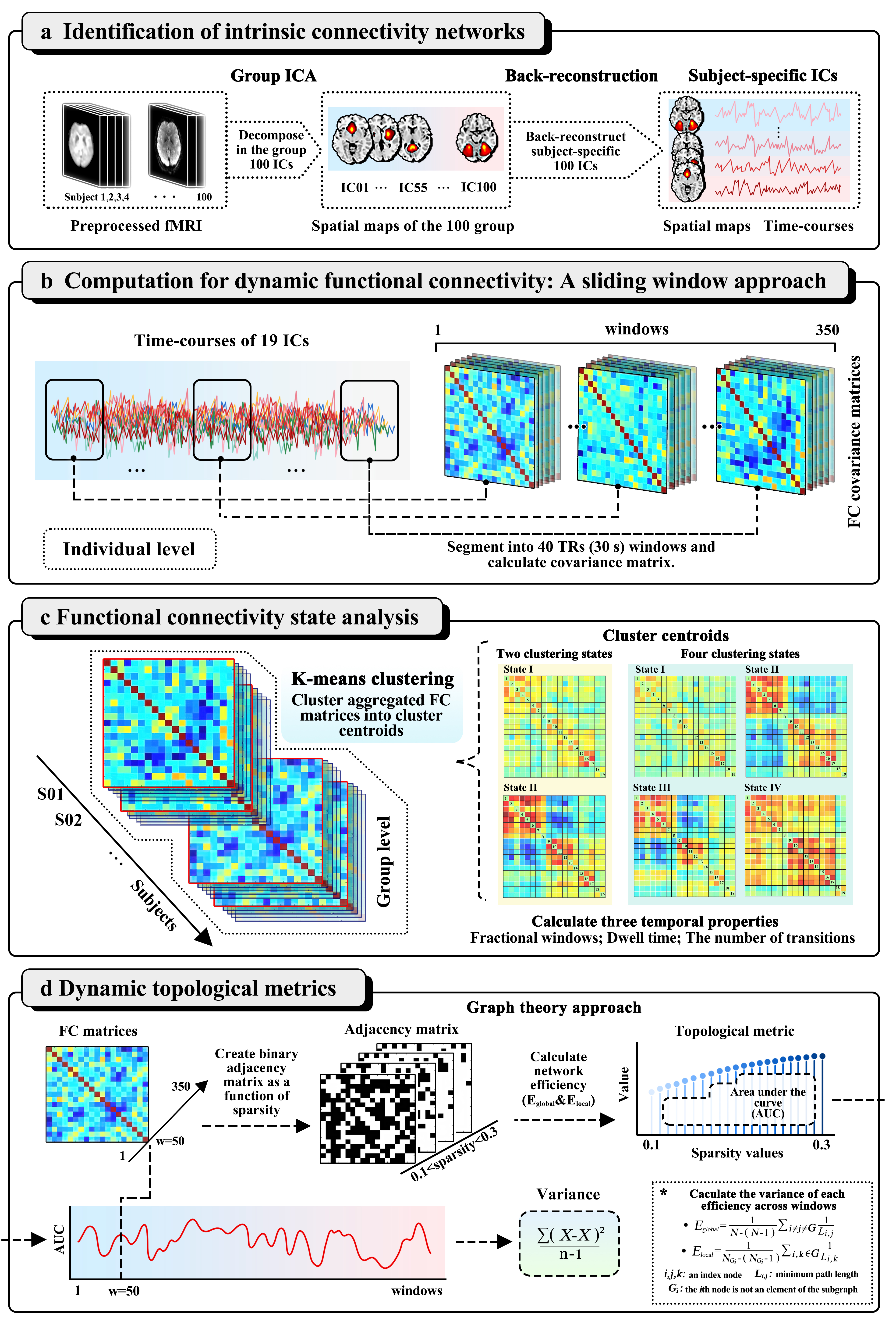
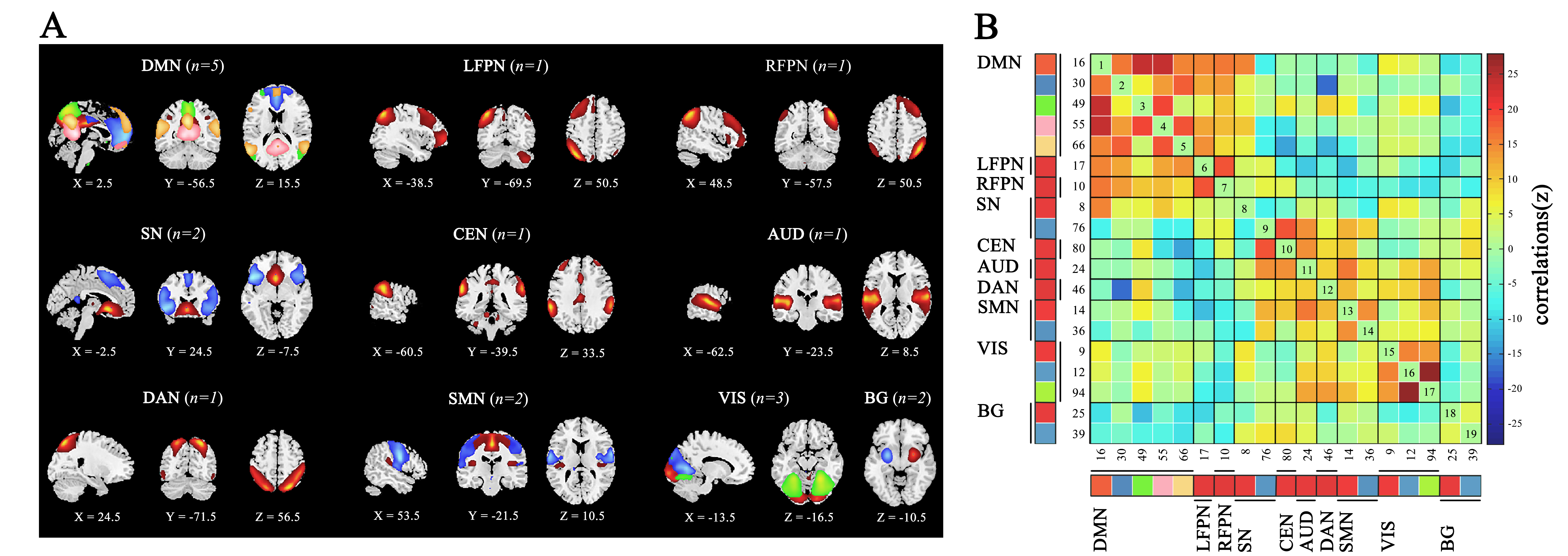
Method: Eighty-seven PD patients, including 29 with FOG and 58 without FOG, and 32 healthy controls were enrolled in this study. Uniquely, we used the MRI protocol with a high temporal-resolution scan, which can provide information on the DFC at a greater temporal resolution (i.e., the repetition time (TR) of MRI scan time is 750 ms) compared with previous studies. Figure 1 illustrates the analysis steps used in this study for DFC. We performed spatial group independent component analysis (ICA) and recognized nineteen meaningful independent components and sorted them into ten functional networks (Figure 2). A sliding-window approach were used to estimate DFC.
Results: Four patterns of structured FC ‘States’ were identified: a frequent and sparsely connected network (State I (50%)), a less-frequent but highly synchronized network (State IV (11%)), and two states with opposite connecting directions between the visual network and the sensorimotor network (positively connected in State II (18%), and negatively connected in State III (22%)) (Figure 3). Compared to the non-FOG group, patients with FOG spent significantly less time in State II, and more time in State III. The longer dwell time in state III was correlated with more severe FOG symptoms. The fractional window of State III tended to correlate to visual-spatial and executive dysfunction in FOG, but not in non-FOG. Fewer transitions between brain states and lower variability in local efficiency were observed in FOG, suggesting a relatively ‘rigid’ brain.
Conclusion: This study highlights how visual network dynamics are related to the presence and severity of FOG in PD patients. These findings can provide new insights into understanding the pathophysiological mechanisms that underly FOG, and may potentially be used to inform a novel neuroimaging marker for FOG in PD.
References: [1] Macht M, Kaussner Y, Moller JC, et al. Predictors of freezing in Parkinson’s disease: a survey of 6,620 patients. Mov Disord 2007;22:953-956.
[2] Kim J, Criaud M, Cho SS, et al. Abnormal intrinsic brain functional network dynamics in Parkinson’s disease. Brain 2017;140:2955-2967.
To cite this abstract in AMA style:
D. Su, L. Ji, Y. Cui, J. Zhou, T. Wu, J. Stoessl, Y. Liu, T. Feng. Altered visual network dynamics associated with freezing of gait in Parkinson’s disease [abstract]. Mov Disord. 2022; 37 (suppl 2). https://www.mdsabstracts.org/abstract/altered-visual-network-dynamics-associated-with-freezing-of-gait-in-parkinsons-disease/. Accessed September 13, 2025.« Back to 2022 International Congress
MDS Abstracts - https://www.mdsabstracts.org/abstract/altered-visual-network-dynamics-associated-with-freezing-of-gait-in-parkinsons-disease/


