Category: Parkinson's Disease: Neurophysiology
Objective: To investigate the correlation between local field potential (LFP) evoked responses, recorded via the DBS lead in Parkinson’s disease patients to stimulation direction
Background: DBS is an established therapy for medically refractory Parkinson’s disease (PD). To obtain optimal effects, the therapeutic brain regions have to be selectively targeted while avoiding regions causing side effects. The advent of directional leads has made this task easier, with many studies showing improved clinical outcomes.1 However, the effect of directional DBS on electrophysiological brain responses has not been investigated.
Method: LFPs were recorded intraoperatively during subthalamic nucleus (STN) implantation in PD patients. Bipolar DBS stimulation was delivered at 10Hz for 30 seconds from the directional contacts on one ring. One of the contacts was used as the cathode, while the other two were used as the anode. For each directional contact as a cathode, a recording was made. The evoked potentials in response to stimulation were calculated by averaging and filtering. Peaks were extracted and analysed. These responses were related to the direction of the cathodic contact, which was determined post-operatively using Lead-DBS. The effect of directional contact was investigated with analysis of variance (ANOVA).
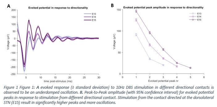
Results: Pilot data (collection is continuing) showed the evoked response to DBS stimulation to be an under-damped oscillation (Figure 1A) in agreement with a previous study.2 When stimulating in direction of the dorsolateral STN, the evoked resonance was observed to have higher peak-to-peak amplitudes and more oscillations (Figure 1B). The ANOVA showed a clear effect of directionality for the amplitude of the first three peaks, present in all directions (F: 396.89; p < 0.0001, F: 655.69; p < 0.0001, F: 412.56; p < 0.0001). A post-hoc analysis confirmed a significant difference between each of the contacts.
Conclusion: Early data shows that DBS stimulation in the STN results in an under-damped oscillating evoked response. Furthermore, stimulation direction is shown to have an effect on peak-to-peak amplitudes and number of peaks, with peaks directed at the dorsolateral motor area of the STN having higher and more peaks. This area has consistently been linked to improved motor outcomes3, indicating the evoked response could be linked to clinical outcomes. However, more research is needed to confirm these findings.
References: 1. Shub A, Bezchlibnyk YB, Smith DA, et al. Clinical Outcomes from Directional Deep Brain Stimulation in Parkinson’s Disease Patients: A Retrospective Analysis (1018). Neurology. 2020;94(15 Supplement). 2. Wiest C, Tinkhauser G, Pogosyan A, et al. Local field potential activity dynamics in response to deep brain stimulation of the subthalamic nucleus in Parkinson’s disease. Neurobiol Dis. 2020;143:105019. doi:10.1016/j.nbd.2020.105019 3. Herzog J, Fietzek U, Hamel W, et al. Most effective stimulation site in subthalamic deep brain stimulation for Parkinson’s disease. Mov Disord. 2004;19(9):1050-1054. doi:10.1002/mds.20056
To cite this abstract in AMA style:
T. van Bogaert, J. Peeters, A. Boogers, P. de Vloo, B. Nuttin, M. Mc Laughlin. Directional deep brain stimulation causes distinct intraoperative LFPs in Parkinson’s patients [abstract]. Mov Disord. 2021; 36 (suppl 1). https://www.mdsabstracts.org/abstract/directional-deep-brain-stimulation-causes-distinct-intraoperative-lfps-in-parkinsons-patients/. Accessed April 26, 2025.« Back to MDS Virtual Congress 2021
MDS Abstracts - https://www.mdsabstracts.org/abstract/directional-deep-brain-stimulation-causes-distinct-intraoperative-lfps-in-parkinsons-patients/

