Category: Parkinson's Disease: Neuroimaging
Objective: We aimed to determine whether microstructural characteristics of key grey matter nuclei would be differentially altered between baseline and two-year timepoints in Parkinson’s disease (PD) compared to controls using diffusion kurtosis imaging (DKI).
Background: DKI may act as a direct, quantitative, and objective measure of tissue microstructure to aid in PD diagnosis and monitoring [1]. However, there are no longitudinal studies of early PD using DKI.
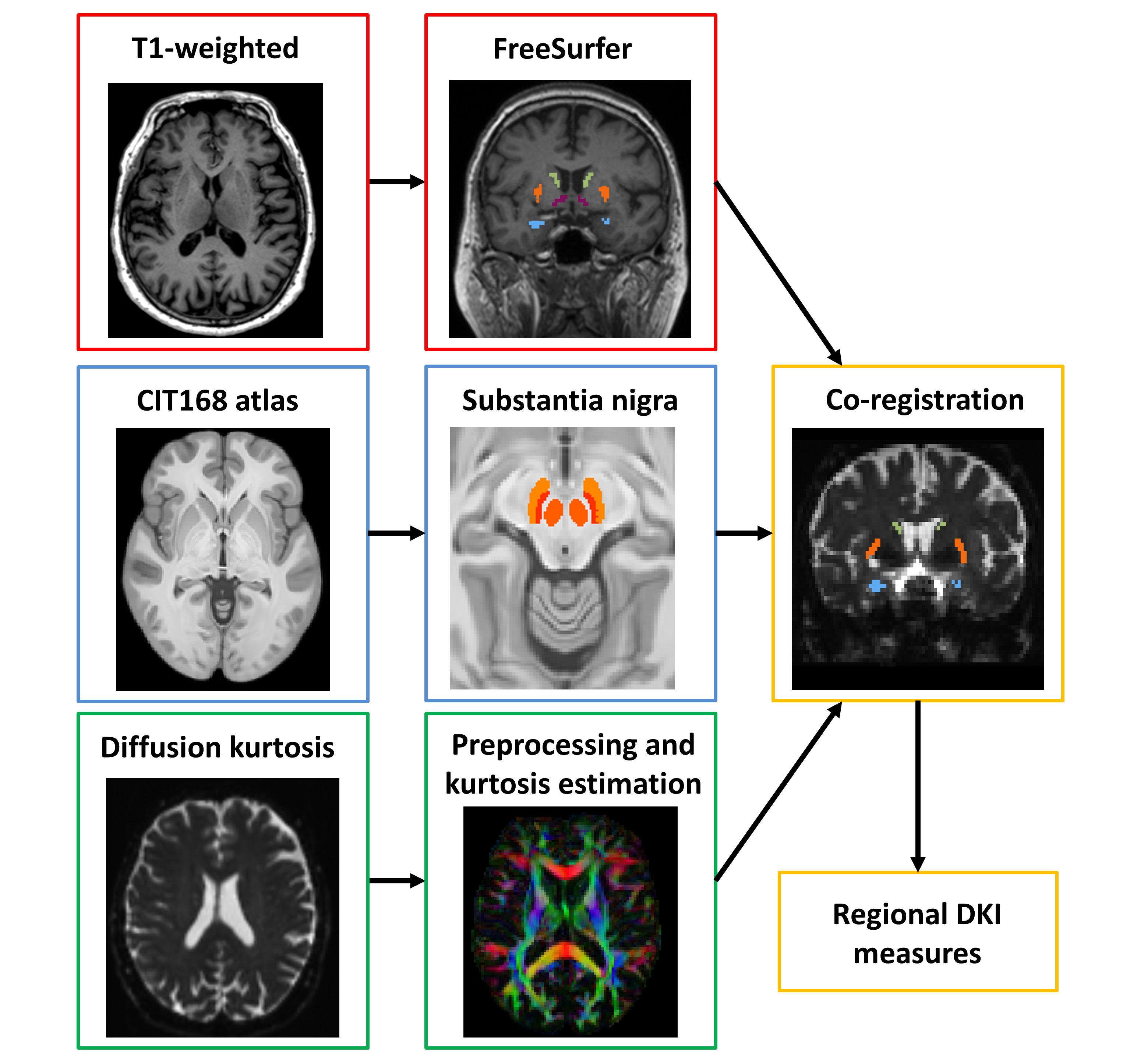
Method: PD patients and healthy controls underwent DKI and clinical assessment at baseline and two-year timepoints. We segmented the substantia nigra (SN) and five other nuclei on MRI images and compared the mean kurtosis (MK) and other DKI indices between groups and over time using repeated measures ANOVA (Figure 1).
Results: The PD group (n=185; aged 67.5±9.1 years; 43% female; MDS-UPDRS-3 20.8±9.8; H&Y 1.7±0.4; disease duration 135.5±100.8 days) and control group (n=77; aged 66.6±8.1 years; 53% female) had an average interval of 2.03±0.24 years between baseline and follow-up.
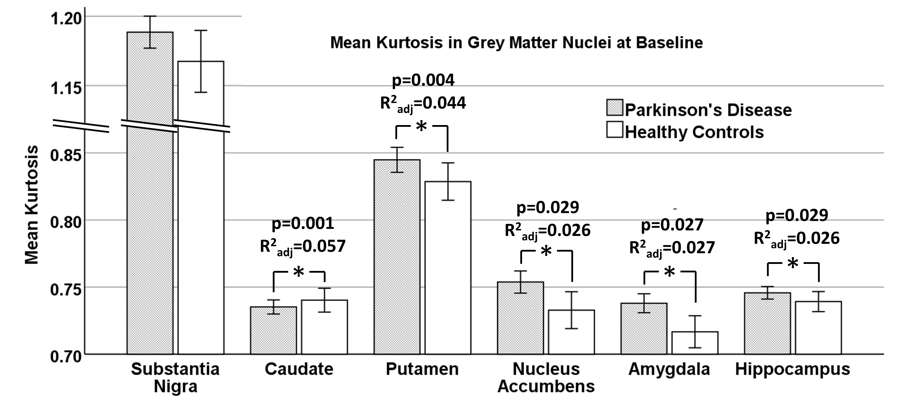
At baseline, MK was higher in PD than controls in the putamen, nucleus accumbens, amygdala and hippocampus when adjusting for age, sex and education but not the SN (R2adj>0.026, p<0.029; Figure 2). In the secondary DKI metrics, the PD group had increased diffusivity, increased kurtosis, lower kurtosis fractional anisotropy (kFA) and no differences in FA across regions (Figure 2). The SN had relatively high kurtosis compared to other ROIs, and the greatest differences between groups were found in the diffusivity of the hippocampus.
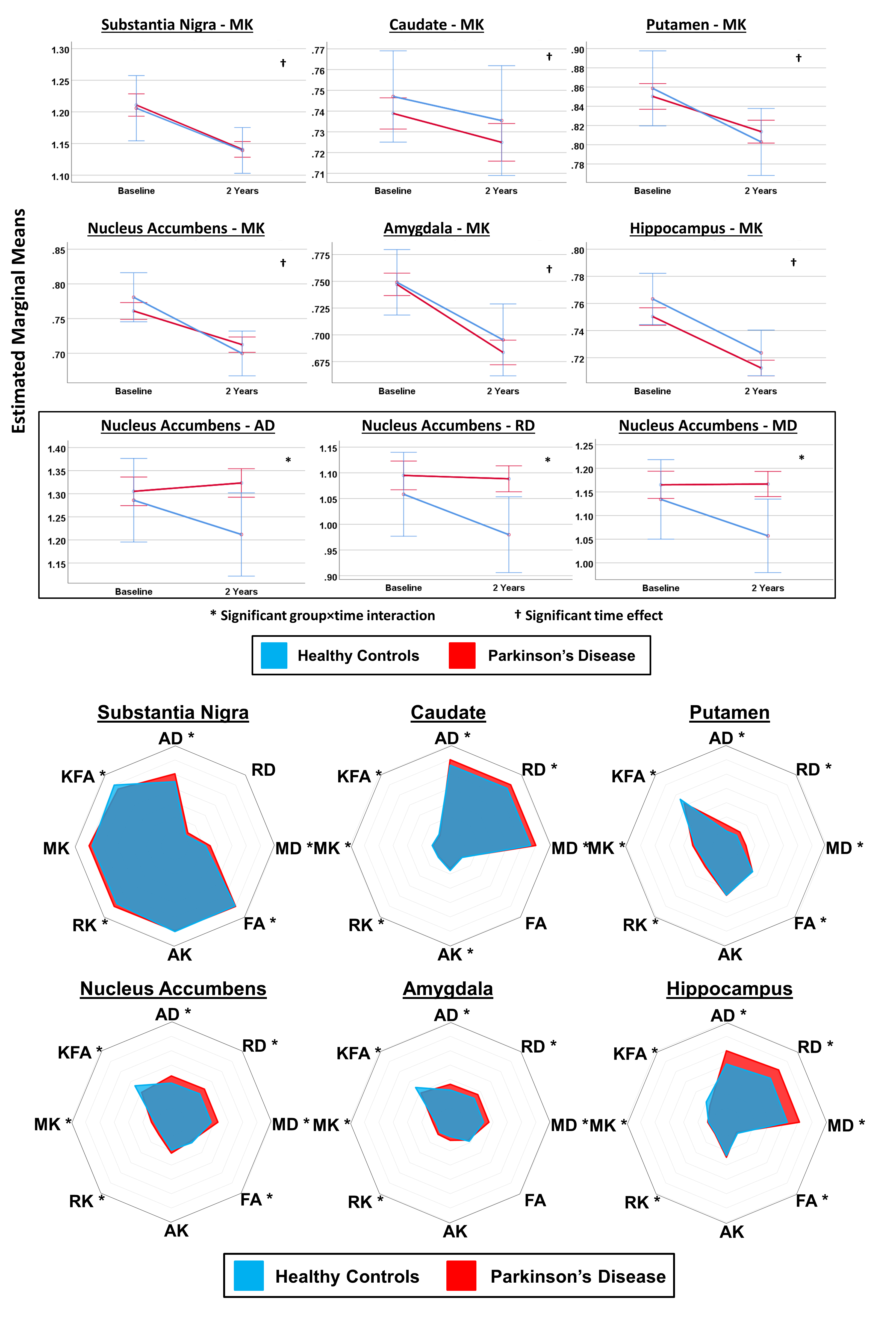
In the longitudinal tests, we found no significant group-time interactions for MK in any region but, for the secondary DKI indices, there were significant group-time interactions for axial, radial and mean diffusivity (p=0.036, 0.032, 0.029, respectively), where diffusivity fell in controls at the expected rate due to ageing but remained static in PD (figure 3).
The two-year change in MK in the amygdala was significantly negatively correlated with the corresponding change in motor performance (r=-0.25, p=0.018).
Worsening of the motor performance was positively correlated with the baseline diffusivity and kurtosis fractional anisotropy (r>0.24, p<0.024).
Conclusion: DKI of specific grey matter structures can be useful to quantitatively monitor PD progression, and may provide prognostic value for deterioration of motor function.
References: 1. Jensen, J.H., et al., Diffusional kurtosis imaging: The quantification of non-gaussian water diffusion by means of magnetic resonance imaging. magnetic resonance in medicine, 2005. 53(6): p. 1432-1440.
To cite this abstract in AMA style:
T. Welton, S. Hartono, Y-C. Shih, W. Lee, SL. Chong, S. Ng, N. Chia, EK. Tan, LL. Chan, L. Tan. Longitudinal Study of Grey Matter Nuclei in Early Parkinson’s Disease using Diffusion Kurtosis Imaging [abstract]. Mov Disord. 2021; 36 (suppl 1). https://www.mdsabstracts.org/abstract/longitudinal-study-of-grey-matter-nuclei-in-early-parkinsons-disease-using-diffusion-kurtosis-imaging/. Accessed February 3, 2026.« Back to MDS Virtual Congress 2021
MDS Abstracts - https://www.mdsabstracts.org/abstract/longitudinal-study-of-grey-matter-nuclei-in-early-parkinsons-disease-using-diffusion-kurtosis-imaging/


