Category: Parkinson's Disease: Neuroimaging
Objective: To investigate the differences in functional connectivity among healthy subjects (HC), patients with REM sleep behaviour disorder (RBD) and levodopa-naïve patients in early stage of Parkinson’s disease (PD).
Background: Dysfunctional resting brain network in PD has previously been described at multiple levels [1, 2]. As RBD is considered a risk factor for neurodegeneration potentially leading to PD, we hypothesized a similar pattern of aberrant functional connectivity albeit with various severity in both disorders.
Method: The functional connectivity was assessed using a data-driven approach with eigenvector centrality (EC) to identify communication nodes playing a central role within the entire brain network. Sixty levodopa-naïve PD patients (20F, mean age 63±(SD)10 years), 57 RBD patients (10F, 65±6y) and 50 HC (12F, age 62±9y) were examined by resting-state functional 3T MRI. While general connectivity was mapped by EC (ReLU correlation), selective connectivity was calculated from seeds derived from the results of a group EC analysis, which implemented GLM with contrast among three groups of subjects using p<0.001 for threshold at peak level and p<0.05 for family-wise error correction at cluster level.
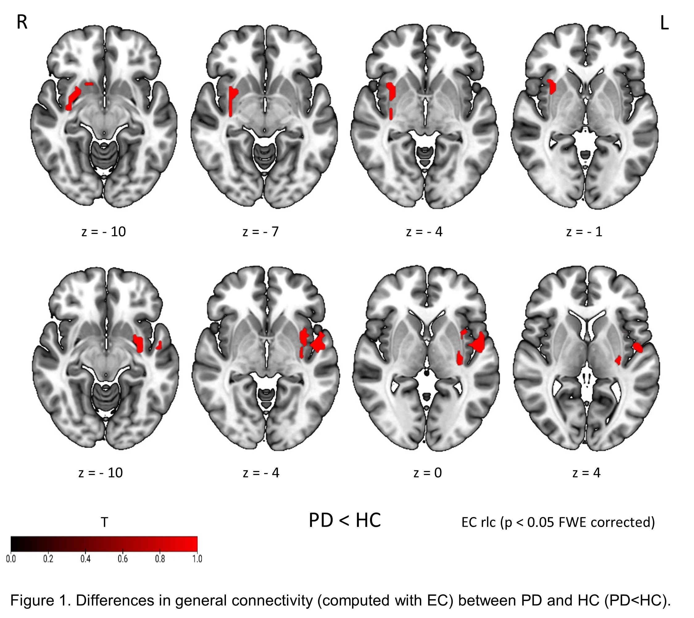
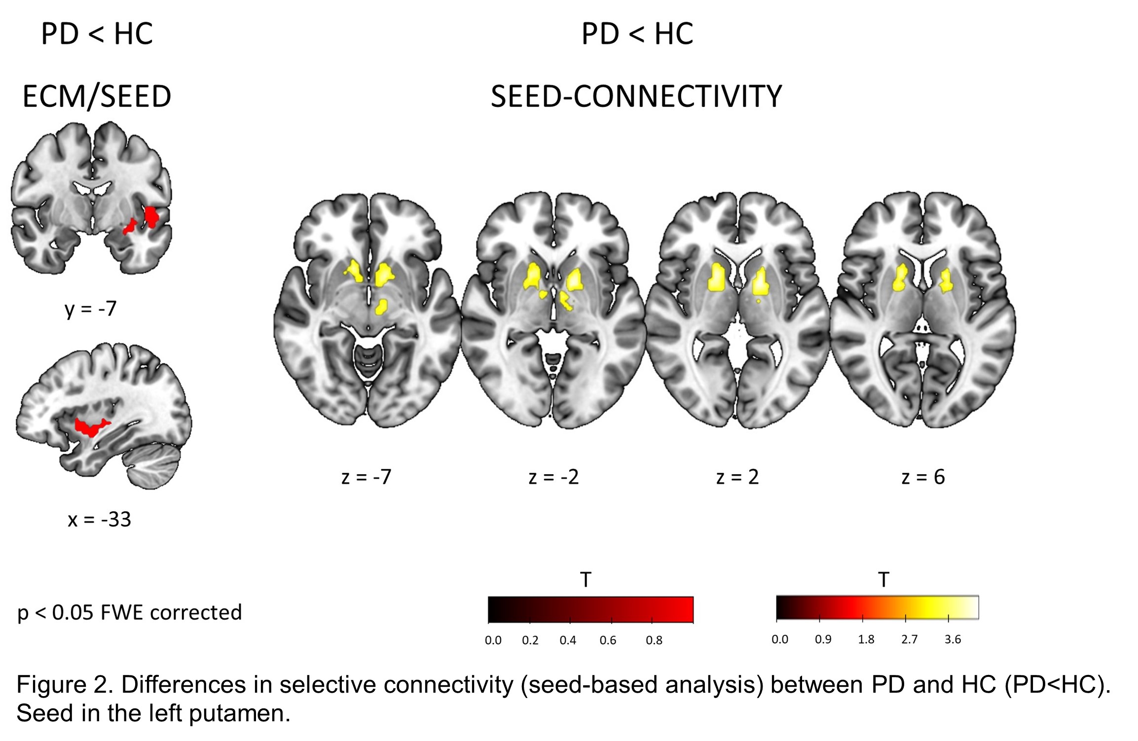
Results: Compared to healthy controls, PD patients showed lower general connectivity (EC) in the insula and putamen bilaterally [figure1]. Analysis of selective connectivity revealed decreased connectivity between the left insula/putamen and ventral basal ganglia bilaterally in the PD than in the HC group [figure2]. However, no differences in connectivity of RBD patients from other groups were detected.
Conclusion: Our results suggest that disconnection affects the basal ganglia already at an early stage of PD. As only levodopa-naïve patients were included, the aberrant resting connectivity reflects the early consequences of neurodegeneration and not the impact of dopaminergic medication which may predominate at later stages [2, 3]. Interestingly, the resting connectivity pattern in RBD was indistinguishable from the pattern in the PD and HC groups which could potentially suggest a subtle network involvement in the brain of RBD patients further supporting this disorder as a pre-stage of PD. Supported by the grant AZV NV19-04-00233.
References: 1. Prodoehl J. et al. Curr Neurol Neurosci Rep. 2014 Jun;14(6):448 2. Ballarini T et al. Sci Rep. 2018 Sep 25;8(1):14328 3. Mueller K, et al. Brain Commun. 2020 Jan 29;2(1):fcaa005
To cite this abstract in AMA style:
P. štofaniková, F. Růžička, K. Mueller, T. Ballarini, E. Růžička, K. šonka, P. Dušek, R. Jech. Dysfunctional resting connectivity in early stage of Parkinson’s disease is indistinguishable from connectivity in REM sleep behaviour disorder [abstract]. Mov Disord. 2021; 36 (suppl 1). https://www.mdsabstracts.org/abstract/dysfunctional-resting-connectivity-in-early-stage-of-parkinsons-disease-is-indistinguishable-from-connectivity-in-rem-sleep-behaviour-disorder/. Accessed February 14, 2026.« Back to MDS Virtual Congress 2021
MDS Abstracts - https://www.mdsabstracts.org/abstract/dysfunctional-resting-connectivity-in-early-stage-of-parkinsons-disease-is-indistinguishable-from-connectivity-in-rem-sleep-behaviour-disorder/


