Category: Parkinson's Disease: Neuroimaging
Objective: To assess longitudinal changes in neuromelanin (NM) content in the substantia nigra compacta (SNc) and its lateral-ventral subregion in Parkinson’s disease (PD).
Background: NM-sensitive MRI (NM-MRI) studies of PD so far have focused on cross-sectional observations of reduced SNc volume or contrast when compared to controls. However, NM dynamics are complex, and NM loss may not occur linearly throughout PD. Both cellular stress and levodopa stimulate neuromelanin synthesis, while neurodegeneration results in NM loss [1-3]. SNc neurodegeneration in PD is also non-uniform, and the lateral-ventral tier of the SNc is the subregion most profoundly damaged in PD [4,5]. We hypothesized that in early, levodopa-treated PD that net NM accumulation, not loss, may occur over time in SNc or its lateral-ventral tier. To investigate this we longitudinally studied NM-MRI contrast measures in these structures.
Method: Twenty-one patients with early to moderate PD [6] were scanned with a 3T Siemens Prisma-fit MRI scanner at baseline and 16 months. MDS Unified Parkinson’s Disease Rating Scale Part III Motor Exam (MDS-UPDRS-III) and levodopa equivalent daily dose (LEDD) were collected. NM-MRI data was acquired using a magnetization transfer (MT) prepared 2D gradient echo pulse sequence [7,8]. MT was measured in both the SNc and lateral-ventral SNc with a highly reproducible, automated method [9,10]. Paired samples t-tests were used to compare baseline and 16-month measurements of MT contrast (MTC) for both SNc and lateral-ventral SNc, LEDD and MDS-UPDRS-III.
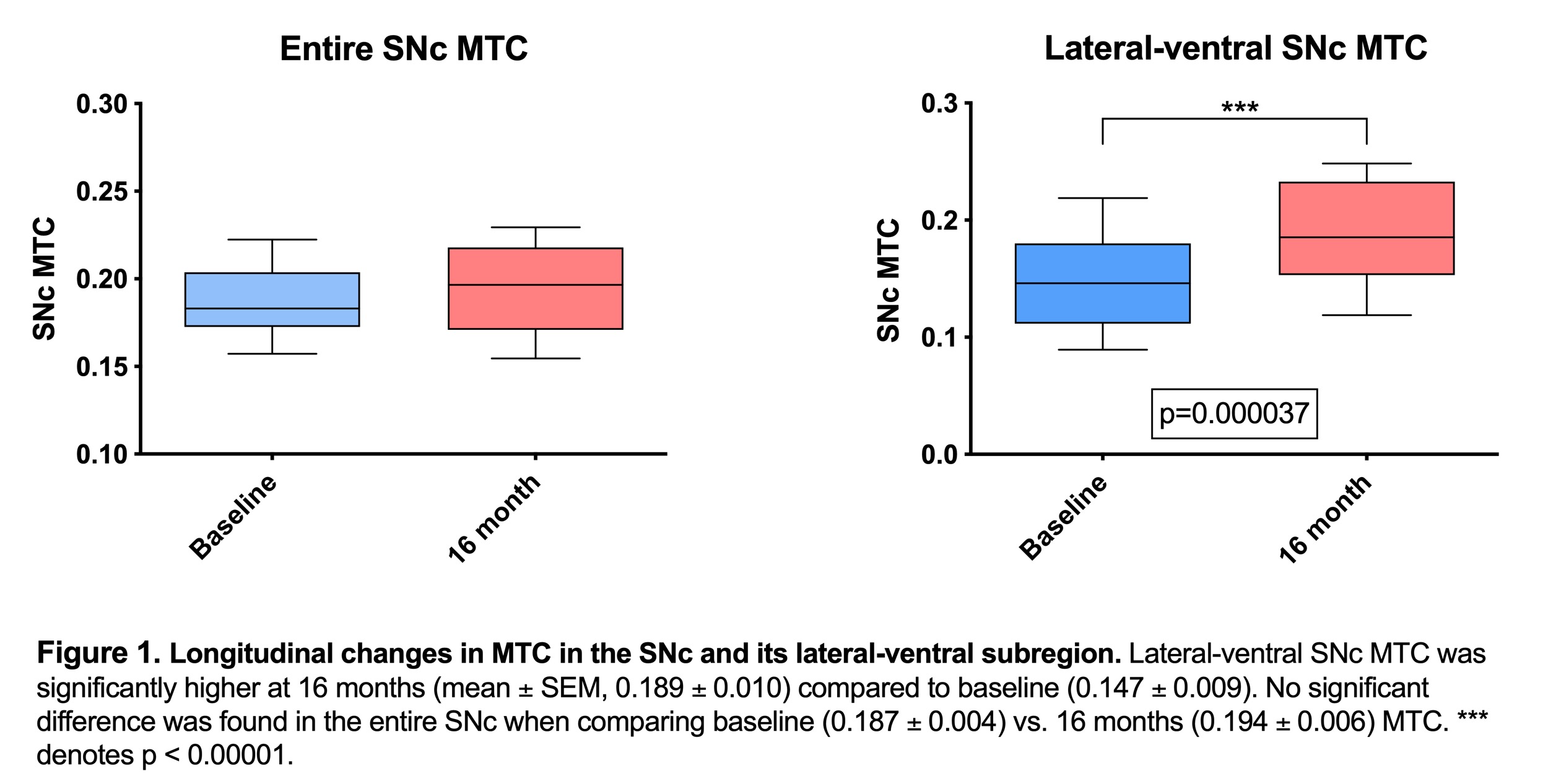
Results: MTC was significantly higher (p=0.000037) in the lateral-ventral SNc at 16 months vs. baseline [Figure 1]. No significant difference (p=0.194) was found between baseline and 16-months MTC in the entire SNc [Figure 1]. LEDD was significantly higher (p=0.007) at 16 months (699.1 ± 69.9) vs. baseline (531.3 ± 62.9). There was no significant difference between baseline (23.6 ± 1.3) and 16-months (24.5 ± 1.7) MDS-UPDRS-III.
Conclusion: Early to moderate PD patients experiencing increasing doses of levodopa with stable motor symptoms exhibit a significant increase in NM-MRI contrast in the lateral-ventral tier of the SNc. These results are consistent with an increase in NM synthesis during this PD stage that may be driven by levodopa exposure and cellular stress. Further studies are needed to investigate neuromelanin dynamics in early stages of PD.
References: 1. Sulzer D, Bogulavsky J, Larsen KE, et al. Neuromelanin biosynthesis is driven by excess cytosolic catecholamines not accumulated by synaptic vesicles. Proceedings of the National Academy of Sciences of the United States of America 2000;97:11869-11874. 2. Zecca L, Zucca FA, Wilms H, Sulzer D. Neuromelanin of the substantia nigra: a neuronal black hole with protective and toxic characteristics. Trends Neurosci 2003;26:578-580. 3. Zhang W, Phillips K, Wielgus AR, et al. Neuromelanin activates microglia and induces degeneration of dopaminergic neurons: implications for progression of Parkinson’s disease. Neurotox Res 2011;19:63-72. 4. Fearnley JM, Lees AJ. Ageing and Parkinson’s disease: substantia nigra regional selectivity. Brain 1991;114:2283 – 2301. 5. Hassler R. Zur Pathologie der Paralysis Agitans und des postenzephalitischen Parkinsonismus. . J Psychol Neurol 1938;48:387-476. 6. Postuma RB, Berg D, Stern M, et al. MDS clinical diagnostic criteria for Parkinson’s disease. Mov Disord 2015;30:1591-1601. 7. Chen X, Huddleston DE, Langley J, et al. Simultaneous imaging of locus coeruleus and substantia nigra with a quantitative neuromelanin MRI approach. Magnetic resonance imaging 2014. 8. Huddleston DE, Langley J, Sedlacik J, Boelmans K, Factor SA, Hu XP. In vivo detection of lateral-ventral tier nigral degeneration in Parkinson’s disease. Human brain mapping 2017;38:2627-2634. 9. He N, Langley J, Huddleston DE, et al. Improved Neuroimaging Atlas of the Dentate Nucleus. Cerebellum 2017;16:951-956. 10. Langley J, Huddleston DE, Liu CJ, Hu X. Reproducibility of locus coeruleus and substantia nigra imaging with neuromelanin sensitive MRI. MAGMA 2017;30:121-125.
To cite this abstract in AMA style:
K. Hwang, J. Langley, N. Kurra, S. Factor, X. Hu, D. Huddleston. Longitudinal changes in nigral neuromelanin in early, levodopa-treated Parkinson’s disease [abstract]. Mov Disord. 2021; 36 (suppl 1). https://www.mdsabstracts.org/abstract/longitudinal-changes-in-nigral-neuromelanin-in-early-levodopa-treated-parkinsons-disease/. Accessed January 13, 2026.« Back to MDS Virtual Congress 2021
MDS Abstracts - https://www.mdsabstracts.org/abstract/longitudinal-changes-in-nigral-neuromelanin-in-early-levodopa-treated-parkinsons-disease/

