Category: Surgical Therapy: Parkinson's Disease
Objective: To identify the patterns of gray matter volume by voxel-based morphometry (VBM) in responders and non-responders with Parkinson’s disease (PD) after deep brain stimulation (DBS).
Background: Outcome predictors such as levodopa-response have been investigated to refine the process of candidate selection in DBS treatment for PD. However, even for the most widely used predictor, there were studies suggesting its results and DBS responses are not congruent. With the advent of data processing technique, pre-operative MRI data is receiving increasing interest in outcome prediction. Though studies suggested structural MRI for prediction of outcome, the results were not consistent and few studies investigated patients received globus pallidus internus (GPi) DBS.
Method: PD patients treated with bilateral subthalamic nucleus (STN)- or GPi-DBS with pre- and post-surgical UPDRS III motor function examination were included. Eligible patients should be absent of obvious cognitive dysfunction. Responders of DBS were defined as having ≥30% improvement in UPDRS III in post-operative med-OFF and DBS-ON state compared to pre-operative med-OFF state. Image preprocessing was conducted using SPM12. After segmentation and normalization, grey matter (GM) maps were compared between groups of patients using a two-sample t test (voxel>30, FDR correction p=0.05 or uncorrected p=0.001).
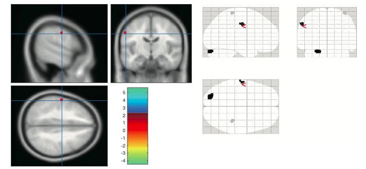
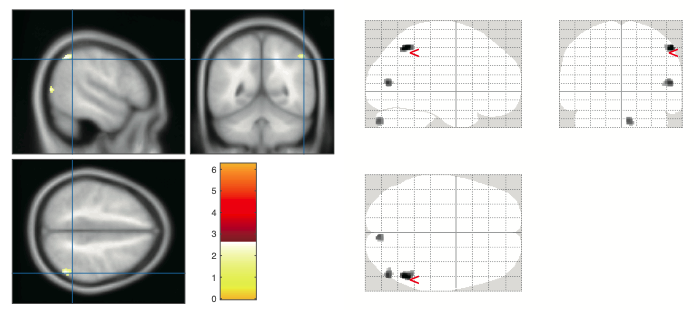
Results: 53 patients (29 in STN-DBS group, 24 in GPi-DBS group) were included in the final analysis. At last follow-up, as assessed with UPDRS III total score, the motor symptoms significantly improved in both STN-DBS group (from 54.4±15.1 to 34.3±11.3, 16 responders, at 6.6±2.3 months) and GPi-DBS group (from 47.5±16.5 to 36.3±14.4, 12 responders, at 7.5±2.9 months). No significant difference was found in other clinical variables between responders and non-responders in each group. In STN-DBS responder group, lower volumewas found in right postcentral gyrus; higher volume was found in regions in left cerebellum and left postcentral gyrus (Figure 1). In GPi-DBS responder group, lower volumes were found in regions in right cerebellum, right middle temporal gyrus and right interior parietal gyrus (Figure 2).
Conclusion: Our data support the notion that the results of pre-operative structural MRI image with voxel-wise comparison could be used as a potential predictor of outcome in the treatment of DBS in PD.
To cite this abstract in AMA style:
Y. Lai, P. Huang, C. Zhang, T.o Wang, B. Sun, D. Li. Differential patterns of gray matter volume in responders and non-responders with Parkinson’s disease after deep brain stimulation [abstract]. Mov Disord. 2020; 35 (suppl 1). https://www.mdsabstracts.org/abstract/differential-patterns-of-gray-matter-volume-in-responders-and-non-responders-with-parkinsons-disease-after-deep-brain-stimulation/. Accessed February 2, 2026.« Back to MDS Virtual Congress 2020
MDS Abstracts - https://www.mdsabstracts.org/abstract/differential-patterns-of-gray-matter-volume-in-responders-and-non-responders-with-parkinsons-disease-after-deep-brain-stimulation/


