Category: Parkinson's Disease: Neurophysiology
Objective: To find the specificity of neuronal responses in the subthalamic nucleus during externally and internally generated voluntary movements in Parkinson’s disease.
Background: The motor control hypothesis proposed that cerebello-thalamocortical (CTC) pathway would dominate during externally-triggered (ET) movements, whereas the basal ganglia (BG)–thalamocortical circuit would show a predominant involvement in internally-guided (IG) movements. Electrophysiological studies showed that the subthalamic nucleus (STN) as a part of basal ganglia is involved in the preparation of both ET and IG movement in Parkinson’s disease [1]. At the same time, neuronal mechanisms of ET and IG motor control remain unclear.
Method: 10 PD patients in an awake state undergoing DBS surgery participated in the study. We performed intraoperative microelectrode recording (MER) of single unit activity in the subthalamic nucleus. On each side, we asked patients to clench their contralateral fists in response to verbal commands and themselves. We performed EMG recording of flexor and extensor muscles as well as acoustic signals of verbal commands simultaneously with MER data. We found and analyzed the characteristics rhythmic cells based on their oscillation scores. We used perievent spectrogram and wavelets to analyze the time-frequency dynamic at the different phases of voluntary movement.
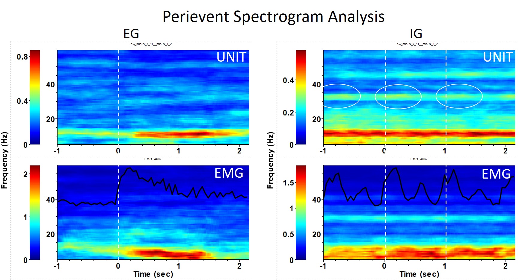
Results: We found that the most typical response of STN cells to both ET and IG movements was the suppression of low beta (or alpha) rhythm from the initiation phase till movement realization. At the same time, we found stabilization of delta and low beta (or alpha) rhythm during holding phase and especially during aftereffect. We also found that in some cases EG and IG movement causes different cell responses. We showed that at the moment of desynchronization of low-beta (or alpha) rhythm, we observed stabilization gamma rhythms during IG movements but not EG movement [figure1].
Conclusion: In summary, our results showed electrophysiological differences in motor signal transmission in the basal ganglia during EG and IG movements in Parkinson’s disease. We suggest that increased “prokinetic” gamma activity during IG movement may be a compensatory mechanism for suppressing of “antikinetic” beta rhythm. The study was funded by the Russian Foundation for Basic Research (19-315-90097 and 18-015-00140).
References: 1 . Purzner, J., Paradiso, G. O., Cunic, D., Saint-Cyr, J. A., Hoque, T., Lozano, A. M., … & Chen, R. (2007). Involvement of the basal ganglia and cerebellar motor pathways in the preparation of self-initiated and externally triggered movements in humans. Journal of Neuroscience, 27(22), 6029-6036.
To cite this abstract in AMA style:
V. Filyushkina, E. Belova, S. Usova, V. Popov, A. Tomskiy, A. Sedov. Rhythmic activity of subthalamic neurons during externally and internally generated movements in Parkinson’s disease [abstract]. Mov Disord. 2020; 35 (suppl 1). https://www.mdsabstracts.org/abstract/rhythmic-activity-of-subthalamic-neurons-during-externally-and-internally-generated-movements-in-parkinsons-disease/. Accessed April 2, 2025.« Back to MDS Virtual Congress 2020
MDS Abstracts - https://www.mdsabstracts.org/abstract/rhythmic-activity-of-subthalamic-neurons-during-externally-and-internally-generated-movements-in-parkinsons-disease/

